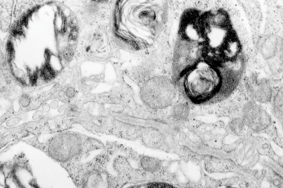
Here is a summary table (already needing to be updated to ferret pix=365; guinea pix=664 with animals being n=61; rat pix=44, n=3: for a new total number of micrographs of 1456), which I could not resist making “pretty”. It does give you an idea of how many electron micrographs have been perused in order to make some statements about whether this protein granule is a part of the regular surfactant machinery or whether it represents something in the way of an “overproduction of protein – hypothetically, surfactant protein A) in a disease state. (I am leaning toward this view). (115 total, 12 species, at least two dozen obscure experimental groups, ages and controls mixed in, three types of processing fixatives, at least three embedding compounds, silver and gold sections, standard uranyl acetate lead citrate staining, and half a dozen different electron microscopes utilized over 30 years, and tissues from at least a dozen different investigators (their materials plus mine). Well over 1000 type II cell images examined in detail.