Monthly Archives: September 2016

Lung lesion: guinea pig alveolar type II cell

As promised, here is another pseudocolored electron micrograph (TEM) that I posted before, this one has all the mitochondria, and the golgi and the RER profiles separated out, in addition to the nucleus, lamellar bodies, and cytoplasm shown before.

I really wanted to add some “teeth” to this to make a joke for halloween…. I might do that, because the blue nucleus looked so much like a “skull” shape, just by chance.

But in seriousness, this lesion of a guinea pig type II cell just didnt show me any proviles of RER that had any kind of striations, layering, or indication that there was any protein organization that might be construed as what is seen in the other guinea pig alveolar type II cells shown in this quest.

Nucleus, green; lamellar bodies, blue; RER, orange; golgi, orange-red; mitochondria, turquoise blue; cytoplasm, pinkish; background and other tissue, pale peach.

4506_M8035_lung_lesion_typeII_pneumocyte_1

Lung lesion: guinea pig alveolar type II cell

This electron micrograph (image) is from a guinea pig lung which was part of a study for vinyl chloride exposure and also given doses of vitamin C.  Gathering tissues, I suspect there was something suspicious that caught our eyes, and a tissue block was taken and marked “lung lesion”. No further diagnosis was given, but in searching for what might be the intracisternal granule in type II cells from guinea pig, ferret, and dog, I found it interesting NOT to find the organized protein in the RER cisternae or in the perinuclear space (often the site of the lamellated protein structure) in alveolar type II cells of this particular lung lesion, even though there were small dilated profiles of RER.  Image below is pseudocolored: nucleus, green; lamellar bodies (of which there are quite a few, and even some in an invagination of the nucleus, blue; cell cytoplasm,  pink; background tissues, orange.  I will pseudocolor the RER and mitochondria as well. There are many free ribosomes, maybe a little more than usual.

4506_M8035_lung_lesion_typeII_pneumocyte

Alveolar proteinosis image: periodicity to the structure

Online there is this image of a lamellar structure which is, I suppose, in the alveolus of some species.  The labels in this publication are not that clear. The author data are: Pulmonary alveolar proteinosis. R. Borie, C. Danel, M-P. Debray, C. Taille, M-C. Dombret, M. Aubier, R. Epaud, B. Crestani European Respiratory Review 2011 20: 98-107; DOI: 10.1183/09059180.00001311, but they reference someone else for producing this micorgraph, but that reference did not show up any images such as this.  So, it is unfortunate because no species, treatment, magnification or other descriptors are provided for this protein-lipid-whorl structure, and it does have the two dense line – central dotted line that the RER profiles in some of the alveolar type II cells posted on this blog, in search of the identity of the protein in those profiles.  It would have been great to know the magnification, and hence the number of nm between the two dense lines of this lamellated alveolar structure.  (used without permission, but credit is given as far as i can determine). Blue arrows point to the central line (inset is from upper left-middle of the lower mag image) and red arrows point to the outer dense lines.  It is too bad I don’t have a micron marker for this image.

lavage_surfactant_image1

Cover submission 2012: MicroDNAs

One more submission for a scientific cover failed but none the less really informative and very interesting.  63 awesome little electron micrographs cut and pasted from data from my friend Jack Griffith, assembled into a collage of images behind a circular recreation of ball and stick DNA.  Love the colors.  This was a pretty guts submission, maybe pushing the envelope for those graphically challenged editors.  haha.

microDNAs_cover_submission

Hamster alveolar type II cell: electron micrograph

Still searching, here, for clues as to what types of organizational patterns can appear in the rough endoplasmic reticulum of alveolar type II cells. It becomes apparent that not all species have these organizational things, the intracisternal bodies, or granules. The hamster comes close, perhaps, and if this is not my imagination, I have highlighted a periodicity (mostly dotted) within a few profiles of RER in the cytoplasm of an alveolar type II pneumocyte.  There are some obvious differences between this organization (if it is not artifact) and the granules that appear in the guinea pig, ferret, and dog type II cells that are noteworthy. 1) if the dotted appearance is real, then it is perpendicular to the long axis of this RER profile, and 2) if this is a surfactant protein organization at all, then it doesn’t demonstrate the same periodicity as the other three species.  This doesn’t mean of course that it is not a surfactant protein, ’cause it likely is. Even at the widest part between rows of “dots” there is probably not 100 nm…  maybe 80 or 90, so that puts the size a little off.  Ribosomes in blue can be used as a reference to approx 20-30 nm diameter.

7287_24716_hamster_alveolar_type_II_cell

Yellow orange body is the RER profile with dots,  purple-blue, ribosomes for comparison in size.  (neg 7287 block 24716 hamster, born 9 1981, presumably female, animal 2 of three, @ 1.5 yr old.

Hunting for what E2 is: Freon 12

Per a recent post  HERE about liquid breathing and E2 and the presence of an emulsified E2 within phagolysosomes seen with TEM, and the tendency for the smaller droplets to coalesce at some point into bigger droplets (judged only by time, since in a matter of 10 days post recovery from liquid breathing E2 for 3 hours) at 17 days recovery, the tiny droplets seen at 5 days of recovery are virtually non-existent.  So I was prompted to figure out the name for E2 (it has been 40 years since these experiments were performed with L. C. Clark, Jr. and there was no way I could remember what the acronyms stood for.

E2 was taken out of production in the 1990s in response to claims that it depleted the ozone layer (I have not researched this so I cannot give an opinion). IS THIS STRUCTURE E2?

Here is a summary, and a link to an interactive chemical image of E2, a list of names (actually a long list of alternative names) and a great ball and stick chemical image… showing both the chlorine and the fluorine atoms (both kind of greenish in color) that I found on wikipedia, along with the black and white diagram.

E2_ball_stick_moleculeE2_ball_line_diagram

dichlorodifluoromethane,
E2
R 12
freon 12
CFC 12 (chloro fluoro carbon halo methane)
Dichlorodifluoromethane
Carbon dichloride difluoride
Dichloro-difluoro-methane
Difluorodichloromethane
Freon 12
R-12
CFC-12
P-12
Propellant 12
Halon 122
Arcton 6
Arcton 12
E940
Fluorocarbon 12
Genetron 12
Refrigerant 12

Acronyms are now anacronisms!

My premise is that we have rendered the acronym as a useless tag, just by sheer number — and incidence they are without meaning anymore. 

for example: ATP  –  adenosine triphosphate, Association of Tennis Professionals, ATP Electronics, Accidental Tech Podcast, Association of Test Publishers, Assistive Technology Professionals, ATP Flight School,  etc etc etc,  so those of you out there who think that it is good for advertising to use an acronym are deluded.  A name and its logo need to have some reference that is clearly obvious, and the acronym alone will get you nowhere.

An acronym is a word or name formed as an abbreviation from the initial components in a phrase or a word, usually individual letters (as in NATO or laser) and sometimes syllables.

An anachronism is a thing belonging or appropriate to a period other than that in which it exists, especially a thing that is conspicuously old-fashioned.

Misleading advertising Kroger ground sirloin burgers: Who else got fooled?

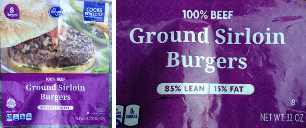
It is a difficult task, to say the least, to keep big companies like Walmart, Kroger, Macy’s, Ford, all the biggies, none is exempt, in line.  Who can do it.  We can’t, except by one phone call at a time.  Seemingly useless gestures, and they probably really are useless, but I am compelled to speak sometimes. THis is one time.  I look at beef as an “occasional” treat, most of you do not, but the data are clear, eating lots of red meat just does contribute to disease risk.  I did get this package of ground beef patty bag, how I got it is sort of irrelevant, but I read the front,  85% lean, 15% fat.  I have seen that add at their meat counter, it seemed reasonable enough.  BUT, look at the label.  Photos of front, of claim for “lean-ness” and compare that with the nutrition label on the back…. WHAT…. the product is not 15% fat at all, but 62% fat…  do the math. (in all due respect and honesty, kroger did call me back with the information i requested — this doesn’t mean that the labeling is not deceptive, just in accordance with the FDA, and it means at least they responded via customer service — it took about a week however)

kroger_ground_beefI called Kroger,  and of course, got the run-a-round.  What is there about this advertising that is fair…. nothing.  You read the front, it gives you percent lean (at least that is what they want you to think) and in fact there are some odd calculations going on here.  What I gleaned (albeit kind of piecemeal… cause I think he was hedging) from the kroger customer service call…. is that they take a piece of meat that is 85% lean, and then add an additional 15% fat….  what…  that is not what this label says, but it is reflected in the nutrition label.  He claimed the labeling was according to FDA USDA guidelines….   if this is so, and I doubt that they are restricted from explaining the calculation on the package so that people like me are not “hoodwinked”…  then we can only laugh at everyone at the FDA and USDA together.  How sad… and you and I pay, we pay for the fraud, we pay for the pain and suffering of our families, we pay for the added health care, and we also pay Kroger profits, and we pay for the salaries of the FDA and USDA staff.  We pay way way too much.

kroger_ground_beef_1and per their own website…. what the heck is  “USER MODIFIED FOOD”

Wondering how many calories are in Ground Round 100% Pure Beef?

Manufactured by Kroger
User modified food.

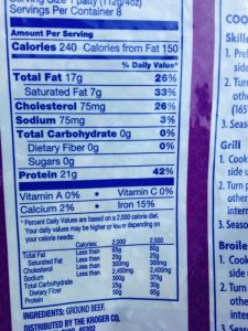
COMMENT: Kroger did call me back with info…. discrepancies lie in the front page units of measure (grams) and the back page calculation units of measure (calories). I should have figured that out myself, nevertheless… the front page is deliberately used to misadvise individuals on the fat content.  fat=9 calories per gram protein=4 calories per gram, which I knew, but didn’t put 2 and 2 together.  DID YOU?

Alveolar macrophage from a mouse that breathed E2 for 3 hours, 5 day recovery

There is an amazing thing that happens to fluorocarbons, in general, when they are taken in by phagocytic cells in the body. It was my observation in the mid 1970s and it remains today, but not well reported: that is — that many (depending upon what species of perfluorochemical it is) types of perfluorocarbon, whether breathed or injected as emulsions, tend to go through a phase of bubble coalescence and then re-emulsification – by the lysosomal enzymes) before they are finally “exhaled” through the cell membranes. I was pretty sure I could tell which perfluorocarbons would be removed in a timely manner from the body by how they behaved in the re-emulsification processes within lysosomes.

Here is an alveolar macrophage from the lung of a mouse which liquid-breathed E2 for three hours and was allowed to recover for 5 days before lung tissue was taken.  See the two types of droplets of E2…. some tiny and some coalescing into bigger droplets. All the dense material around the droplets i believe to be lysosomal enzymes.   This image below is a crop (unfortunately the original print i marked all over and couldn’t clean, so this is a rescan of the negative… i don’t have a wet darkroom any more) it could be printed  again, and scanned, and pseudocolored (i might do it ?? to make a great poster in addition to being a useful image for studying.  (unretouched except for contrast enhancement of the scanned negative – neg 1340 block 4835 swiss albino mouse 16.15g, 3 hrs liquid breathing five days recovery, right mid lobe tissue block, 1-30-1975, fixed in modified Karnovsky’s and osmium. There are literally hundreds and hundreds of little E2 droplets and a few big ones.

1340_4835_E2_3hr_5d_rec_alveolar_macrophage