Monthly Archives: June 2018

FORM FOLLOWS FUNCTION? mitochondrial shape follows function!

Beginning with a general truism – form follows function (a 20th century design mantra coined by Louis Sullivan originally as “form ever follows function”) is so far reaching as to make us marvel that it took so long for biologists, chemists, architects, etc, to write it down. Nature wrote that script 3,900,000,000 years ago.

To be surprised that examining the cell in detail (in this case speaking about mitochondria) is naive. It has been obvious since the invention of the electron microscope that mitochondria in different tissues look different. Before that, not much was known about these little tiny objects in the cell, or the squiggly things running around cells in culture in the early time lapse microscopy, as they are just smaller in size than the light microscope could resolve.

So how marvelous it is to know that when you look at a mitochondrion you are “seeing” the biological processes. Read this particular publication on the shape of cristae in mitochondria, which seems to come from a person who can “visualize the 3D as well as understand the science“.  This means that of the tens of thousands of mitochondria that I have photographed that each one tells its own story, whether it was leaning toward oxidative phosphorylation, or detoxing reactive oxygen species, or moving the cell into apoptosis, or maybe preparing for mitochondrial fission or fusion, depending upon levels of stress, or getting ready to participate in autophagy.  It could be moving energy out of the cell, it could be importing necessary proteins from the cytoplasm, or it could be reproducing its own distinctive mDNA, or transcribing and translating proteins on its own mitoribosomes.

So there is a glich…  not all of these events show molecules that are resolvable with the electron microscopy either.

Shape (elongation or rounding) of the whole organelle, or widening of just segments of the whole organelle has meaning (visualize the bulge of a rat in a snake). The shapeso that cristae assume are clues to activities as well; whether tubular, round, triangular, with wide cristae junctions or narrow, dense matrix, numerous mitoribosomes or few, numbers of intramitochondrial granules and more that I don’t know about. The relative volume density of the matrix is also important. It is difficult to pinpoint mDNA, or even mitoribosomes… they are not easy to find like those cytoplasmic ribosomes neatly coiled on mRNA or attached to ER.

Here is a mitochondrion from which one can generalize: it is elongated (mitochondrial stress); it has narrow cristae near the outer mitochondrial membrane (hairpin bends in cristae signals lots of ATP synthase, there is an area of low cristae frequency and greater matrix, and there are some rounded cristae near the center.  The cristae junctions are for the most part are quite narrow.   Mitochondrion blue, ER (both smooth and rough vesicles greatly enlarged in this Gclc ko NAC rescued 50 day old mouse (#5) neg 18411 block 78375s liver mitochondrion (blue). Aside from two different types of cristae, within and elongated, and very narrow crista junctions, there are other features of this portion of an hepatocyte that are interesting, and one is the vesicle-within-a-vesicle formation of the rougn ER and also the consistent separation of ribobomes on those membranes into little groups of 7-10 ribosomes (almost like a signature) with space between the groups… (more form follows function) (orange)
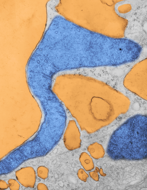

Publication above mentions that folded (tightly) cristae are working cristae, where as those that are spread apart are not functioning well. Mentioning, as i have seen in othe rpublications, that ATP synthase dimerizes or oligomerizes along the sharp curvatures of cristae. Mitochondrion above shows very tight cristae, with hairpin turns, thus one might suppose that ATP synthase is well ordered and working overtime in the Gclc ko mouse model rescued with NAC.

For what it is worth (for me it was worth much) here is a pdf file that some of you may be able to read and find helpful. For me the help came in this paragraph ”
The high proportion of cases reported among these populations suggests a genetic susceptibility for LP. This idea is further supported by cases of familial LP, reported in 1 to 4.3% of childhood LP series (Kanwar and De, 2010; Nanda et al., 2001). However, familial cases have not been reported in all series, and specific antigens have yet to be determined. Pathogenicity of LP lesions involves the autoimmune-mediated lysis of basal keratinocytes by CD8+ lymphocytes, though definitive etiological triggers are still unknown. Case reports in both adults and children have shown an association between LP and the following: chronic liver diseases such as chronic active hepatitis (particularly hepatitis C) and primary biliary cirrhosis; complication of hepatitis B vaccination; viral and bacterial antigens; trauma (via the Koebner phenomenon); metal ions; medications; and a variety of autoimmune diseases such as autoimmune thyroiditis, myasthenia gravis, alopecia areata, vitiligo, thymoma, and autoimmune polyendocrinopathy (Luis-Montoya et al., 2005; Pandhi et al., 2014).  IN PARTICULAR  linking METAL ions to this disease is what I am thinking happened in my case, the metal ions as chromatic agenets in the thinset mortar and the grout that I used on my basement floor. Chromium and cobalt, and hopefully no cadmium, but plenty of other metal elements, and also including the silica, known to cause respiratory disease.

This particular paper suggests that corticosteroids are beneficial, but many sites and publications do not give steroids much credit for shortening the duration of the disease, which can be years. I for one wasnt helped by topical steroid cream (betamethasone was the only one prescribed for me, outside of a very outdated half used tiny sample tube of topicort none others were used).

Next chapter begins a fun 11 or 12 years making ceramic tile collages, montages, outdoor pots (big big ones) and laying 35 thousand tesserae on my basement floor.  Little by little over that span i began to notice a dry cough.  Not that bad, but bothersome.

Case Report: lichen planus – which hypersensitivity? Chapter 2.

[aside]I dont mind posting the names of 5 MDs, none of whom were helpful. Dr Wones, Dr. Zubritsky, Dr. Carter, and Dr. S…(from the middle east) cant remember his name, and the pathologist Dr. Mutasim.  None knew anything about this disease, nor were helpful in any way, in fact Dr. S was downright derogatory, showing complete ignorance about how great the impact of environmental stimuli can be on diseases in general…that is pretty much an old-school mentality, maybe some “updated” information would benefit his overall knowledge of diseases. One sent me a link to a website about lichen planus which only had the most superficial information, totally useless. I guess he thought he was being helpful.

BTW i think that i have had the tendency for lichen planus for many years so what caused the explosion of lesions is really what I want to know.

Several kinds of hypersensitivity have been described (more will likely be found). TYPES I to IV.

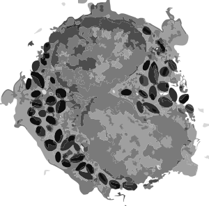
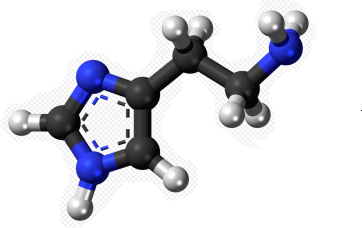
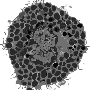
TYPE I -Immediate hypersensitivity happens fast. CD4 T (thymic lymphocyt) cells are involved in adaptive immunity (to pathogens), autoimmunity, asthma, allergic and tumor immunity. Naive CD4 T cells may differentiate into one of several lineages of thymocyte helper cells, (Th1, Th2, Th17, iTreg – about which I know nothing). After the first exposure to the allergen, the CD4 T cells are “sensitized” and immediately effect a response by releasing antibodies (immunoglobulin E (IgE))  which signal mast cells and basophils (with high affinity IgE receptors on their cell membranes and loaded to the hilt with storage granules) to dump out vasoactive amines, e.g. histamine. These chemicals then affect surrounding cells, mainly causing blood vessel dilation and muscle contraction, and cause the inflammation by increasing the permeability of the capillaries to white blood cells and some proteins (thus redness swelling and itching).  Histamine (one of the vasoactive amines) is a cute little molecule (right)(thankUwikipedia). Basophils (left) and eosinophils (center) are cute too (I vectorized two transmission electron micrographs here).

Delayed hypersensitivity is just that, delayed onset. Appearing several hours, days or continuing to appear a week or so after contact with the antigen and depending upon the dose of the antigen. For example poison ivy appearance (at least in my case) comes up between 2 days and a week after exposure, sometimes showing the secondary contact point of the original molecules of urushiol being where two skin surfaces meet..the original spot depositing a lesser amount of urushiol at the point of contact.  While i have never read about it, I do believe there can be systemic poison ivy, for instance when the neighbor weed-wacks their yard full of poison ivy and the droplets of urushiol are aerosolized and breathed.  Would be fun to search that route of exposure (in the literature, not as a test subject).

Delayed hypersensitivity is not antibody dependent, but is a cell-mediated response.


Case Report: lichen planus – is chromium an issue? Chapter 1.

I am going to document what I am thinking about while keeping my hands off the itchy places (not so LOL) to try to figure out what environmental+immune conjunction caused this to surface in my otherwise really healthy 74 year old body.

I don’t know if it will help anyone else in the search for cause, but hopefully it will, and it will allow me do write a history of my own experience (noting everthing I can remember and blaming no one and no thing or even being disparaging..well except maybe a little to the MDs that we depend upon to help us through diseases..and who (in my case) did “not much”. Ha Ha.

I am also going to encourage anyone out there to write a similar history, and though I don’t take comments on this website because so many unscrupulous individuals want to spam and skim and link for their own advantage instead of participating in meaningful discussion. My email can be discovered from the very first entry on this blog, way back when!!, and from one of those websites you can contact me if you want to contribute a long history. My goal is to treat this exactly like a manuscript, and submit it at some point to a scientific journal.

Starting at the beginning (not the beginning of my life obviously, that was 1943) but of contact dermatitis which happened to the best of my recollection in 1968 or 1969, a new time resident of cincinnati, and during a frisby throwing game in burnet woods.  If you are from cincinnati you know burnet woods is a primitive wooded area, small, more a park with a small lake and lots and lots of poison ivy.  I didn’t really know what poison ivy looked like, but in my leather sandals (imported) apparently I ground some poison ivy oil and got a really bad case of it from that afternoon.  I mention it because of the inflammatory process of the poison ivy itself, but back then, leather, and imported leather was tanned with some pretty harsh chemicals, including chromium.  Every time (which was probably one or two more times) I wore the sandals i re-got poison ivy.  Chromium is the key word. Urushiol is the next key word as it is the oil in poison ivy that causes the allergic dermatitis. HERE is a lay website for some important information. A host of things can cause allergic dermatitis; food, cosmetics, metals (i could never wear stainless, nickel plated or silver earrings) and it also can be caused by paints and mortars and cement and grout.

That the exposure on my feet was to 1) several unknown tanning compounds, 2)as well as the urushiol (which causes a T-lymphocyte response), what appears to me to be a really good combination for an adjuvant effect, causing an senhanced response.

I did get poison ivy pretty seriously in 1975, but to my knowledge there was no other environmental exposure at that time. (except possibly, chromium in silk screen paint which i used a lot of (and probably still have some in my basement… SKRINK brand)..greens and blues.  Direct contact of Cr(VI) compounds with intact skin can induce chromium dermatitis or sensitization. and a government document on chromium toxicity.
So chromium has been a part of my life for a long time, though maybe not in a good way.

Fast forward many years, to “fun with polymer clay” when in the 1980s i used lots of FIMO, and likely all colors, though I cannot distinctly remember which colors i used most, but very definitely there was blue, and likely green.  I used enough of that early FIMO to produce a contact dermatitis in the palms of my hands, little bumps, not terribly itchy that i can remember, but enough to make me “wake up” and know that polymer clay in that formulation was a contact allergen for me.  And fast forward another 30 years and it is back to polymer clay again, not too careful this trip around, but no contact dermatitis on my hands (the clay does stick to hands…. especially with some pigments,  black clay and red polymer clay are particularly noticeable, but I don’t doubt for a second that they all stick.

The take home message is that we do get exposures…to many things…. not all of which our bodies treat as benign.


Mitochondrial cristae junctions and pores?

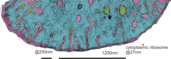
Searching through micrographs to determine whether or not i can visualize a mitochondrial pore or crista junction.  Here is a cutout of a border of a mitochondrion which looked to have several adjacent and more or less evenly spaced cristae along one outer mitochondrial membrane. None of these exposes a clear connection where an inner mitochondrial membrane space should appear. Also of note (in this experimental set of animals) there tends to be a narrowing at the base of the cristae, and a little ballooning of the remainder of the cristae bring. This might be a manifestation of the cristae junction. In a stretch of outer mitochondrial membrane about 475nm in distance there were 6 areas where the former and the cristae membranes meet.

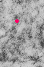
In this particular mouse liver electron micrograph the animal was a hepatocyte specific ko for GCLC, specifically negative 18408 block 78375 wc/ii animal#5 and 50 days old. red dot=@27nm (one cytoplasmic ribosome) used as a micrometer.

mitochondria liver electron micrograph cristae junction
Looking at the matrix of this mitochondrion several densities appear which may be too small of mitoribosomes.


Another mitochondrion which i examined and pseudocolored the same those above, has about 1 cristae membrane junction pore per 250nm or so counted over a stretch about five times that long. Clearly, physical state (these KO mice) nutrition, gender, cell type and function which the mitochondrion is supporting.  Green is the inter cristae membrane space,  pink is the outer and inner mitochondrial membranes, blu background is the mitochondrial matrix. The tissue from which these sections were made was a ko, (conditional KO of GCLC in liver).