Just looking for the myriad features that should show up in mitochondrial ultrastructure. THis isolated mitochondrion is from a wild type mouse post natal day 14, liver. It does show areas where cristae membranes (inner mitochondrial membrane) and outer mitochondrial membranes create the inner mitochondrial membrane space (green) and the membranes themselves (pink) and the matrix (blue). Within the texture of the matrix it is tempting to try to find circular mDNA and mitoribosomes and there are hints of them here but nothing really obvious shows up. Trying to find some difference in inner mitochondrial membrane density which would indicate the crista junction is pretty disappointing as well. Not all structures named, or presumed to be real, can be found in routine micrographs. There is one little circular structure in the middle of the matrix which I might well have included as cristae membrane. 
Monthly Archives: May 2018
Crista junctions in mitochondria
I am trying to visualize crista junctions in mitochondria in the liver (mammalian). These might be visible here, in a KO mouse which has increased oxidative stress (previous posts). It looks to me like there is a small consistent area at the place around cristae approaching the outer mitochondrial membrane that looks “different”? Find the 100nm markers (vertical at the edge of a crista and horizontal by the red dot over on the right hand side) are an approximate marker for the size of a cytoplasmic ribosome (27nm) and calculation for 100nm from that.
Cristae in this particular mitochondrion and many others show an increase in the amount of matrix space and a more vesicular type of cristae, and some times have cristae inclusions.
Handing off the blame!!
Per Rosanne on the news this 30th day of May, I heard on NPR one of the best retorts to a comment attempting to reassign blame for a racist comment from self to Ambien……. Reply from the drug company was spot on and i am sure it will become a classic quote. It went something like this….
“Medicines have many side effects…. racism is not one of them”
What part of your brain chooses your partner for procreation?
Sexual selection in humans — wikipedia has a short general article… I would have thought that this topic would be flooded with blogs and articles…. not so.
I dont have a single sibling who didnt marry a “slower, less anxious, low key, hands off” mentality…. it was great for the species…. but awfully hard on the parental relationships… haha… i laugh about that paradox….. for me it seems very obvious that our animal brains rule… !!! biololgy just is determined to even out the extremes.
Mitochondrial ultrastructure
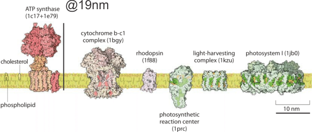
It is a little difficult to get the whole picture of mitochondrial ultrastructure together in one place and in one image. The the large and complex groups of proteins like ATP synthase, or mDNA and mitoribosomes and the proteins for energy conversion, or calcium storage, ion transport, proteins that are involved in cell growth, division and apoptosis, as well as the basic mitochondrial shape and volume density of and shape of cristae and the amounts of mitochondrial matrix, and many other things of which I am sure i am not aware….are difficult to sort out. Ultrastructural aspects related to these functions are not well understood, and at best poorly diagrammed. I have found one website (biology by the numbers) which does do a great job of labeling size and giving measurements of some aspects of mitochondria, but of course not all, and not neatly organized. There really probably is not going to be much consensus about these variations in mitochondrial ultrastructure because the influence of tissue, cell, metabolic state, types of fixation, the embedment, as well as many variations in scope mag, deliberate manipulation of the images, poorly kept records, presumptions, and the list is endless. So the question is how does one go about diagramming (or imaging) the most educational presentation about the mitochondrion. A really perplexing structure is the “pore” or cristae junctions.
Add to that list of variables, the long list of diseases where mitochondrial shape is found and the concept of imaging the “perfect educational” mitochondrion becomes more difficult. So trying to find a great diagram came about because of an inclusion that I found in the cristae of some of my own micrographs.
It would just be nice to see these structures ( 5-10nm diameter) in the literature with the normal ultrastructure of the mitochondrion, which is not really that well documented for standard TEM images though there are some nice molecular diagrams of membrane proteins.
The round electron dense inclusions are unknown (in a crista space)(in these not-perfect micrographs). It is possible that in the bends of a couple of cristae, there are densities right at the bends which would seem to be identifiable. Looking at the blue dots in the top image below, right at the curvature of a crista–one might see those dots as ATP synthase (blue dots are given at the approximate size that ATP synthase should be ( “biologybythenumbers” diagram (thank you for that) (bottom image). Also, separate, larger orange dots could be mitoribosomes which reportedly are just smaller than cytoplasmic ribosomes (picture as red dots (taken from the same micrograph as seen on the left). A second reason for working on mitochondrial ultrastructure is to try to figure out what the electron dense (and homogeneous round) protein is within this crista space)
Looking back at Gclc wc/ii mouse mitochondria
Quoting from Chen, et al, “Hepatocyte-specific Gclc Deletion Leads to Rapid Onset of Steatosis with Mitochondrial Injury and Liver Failure” ……a gene essential for GSH synthesis was disrupted by engineerig the albumin-cyclization recombination (Alb-Cre) transgene to disrupt the Gclc gene specifically in hepatocytes and deletion
within the Gclc gene was mostly complete at postnatal day 14, and GCLC protein nearly depleted at postnatal day 21. GSH depleted in weeks 2 – 3.
Mitochondrial loss of GSH was less severe than cytoplsmic loss but ultrastructural changes in mitochondrial morphology were quite prominent and explanations for those changes (at least in part) were not known at the time. Mitochondria had greatly reduced cristae membrane surface and very much increased matrix. In addition, a ballooning of some portions of the mitochondria “distorted” parallel cristae as if being squeezed to one end or the other of the mitochondrion.
These alterations were accompanied by striking decreases in mitochondrial function in vitro.
Two publications (Daves et al 2011, Daves et al 2012), might shed some light on the shape of cristae (that is their lack of a traditional cristae shape) and possibly relevant to the ballooned matrix of the mitochondria in KO mice. This is regarding the ability of ATP synthase dimers to “bend” the cristae membrane at an angle and being responsible for the acute angle on the edges of flattened cristae, as they are located on ridge area of cristae. The abrupt curvature is classic for mitochondrial cristae particularly in heart muscle, and skeletal muscle, but also in liver, and less prevalent in mitochondrial cristae morphology some other tissues. Also seen within cristae are small round pickets of a homogenous relatively electron dense protein. No explanation for the latter have been found yet.
Humanization kit
Metal? cristae inclusions in mitochondria
 I found a paper that showed a TEM of a dense round intracrista inclusion that had some similarities to what is seen in the Gclcwc/ii hepatocyte specific KO mouse at 50 post natal days. Paper is Characterization of Intracellular Inclusions in the Urothelium of Mice Exposed to Inorganic Arsenic Toxicological Sciences 137(1), 2013 by Puttappa Dodmane et al. I cut and pasted part of one of their electron micrographs of a mitochondrion with such an intracristae inclusion (right side of picture) next to what is seen in the KO mouse (my picture, left side of image).
Mitochondrial and ER inclusions, and nuclear invaginations in Gclc hepatocyte specific KO mice
Hmm. Is there a possibility that the inclusions in the inter-cristae space of mitochondria, the inclusions sometimes seen in nuclei (probably invaginations), and the iron-like dense inclusions in the ER hepatocyte cytoplasm, are linked by enzymes that are involved in the maturation of cellular Fe/S proteins for which mitochondria are important. These images are from Gclc hepatocyte specific KO mice, one image (with the nuclear invagination with iron-like spicules upper left) came from a KO that was rescued with NAC the other two micrographs are from wc/ii mice, unrescued, at day 50.
What kind of diagram is this? mitochondrial cristae
I find that diagrams and illustrations for science are so often NOT GOOD, NOT EXPLANATORY, and are TOTALLY CONFUSING. Here is an example. It is from a very nice article, (Biochimica et Biophysica Acta 1793 (2009) 5–19), but the diagrams of the arrangement of cristae just doesn’t make visual sense. It is an injustice to readers to create confusing images and put them up as learning aids. THis diagram misuses the shading when it creates a look of 3dimsnsions…. it makes no visual sense in any of the three drawings in this figure. To create shading on one portion of a diagram (e.g. the middle figure) which in this case was the intercristae space, which is called the intermembrane space, and NOT make the shading equivalent on the mitochondrial matrix is just careless, and misleading. The lower diagram is beyond deciphering…. it looks like there are three orange fingers poking up, from a space with holes. Nothing resembling anything that is shown with real TEM images. So sad. It wastes time and sends wrong information.


