Monthly Archives: July 2018

Verge of a Dream: Remembering

We don’t need anything new.
Like an updated picture,
you in my arms,
that dress passed on to
your sister. And that
you would stay is gone
too. We didn’t need to
talk further. Stopping
in china town for dinner.
You on my arm,
the late bus passed on by,
like clouds that you made
disappear.
We never grew older,
You had your arm around
me, over my shoulder.
Heroes honored
this weekend.
There
is nothing to change,
I just spend time
in remembering,
how the words that I’m
yours are gone too.

RLB 1 16 2008

Verge of a Dream: An end to all longing

Lets say we rode to
Mount Vernon and
Contemplated the river
Noted the way that stuart
Clouded the picture or
Perhaps was
Floating a ditty.
Lets say our thoughts
Came together
One was a resin
The other a catalyst
Ingraining our senses with
Flowers and gardens that
Bound from the past
On an historical journey
And tomorrow
Awoke to an early
Sun on a summers day
An exuberant display
And an end to
All longing

RLB  1-24-2008

Identifying and diagramming mitochondrial proteins

This group did, in my opinion, a reasonably good job of modeling mitochondrial membrane proteins in a way that utilizes all the information (likely) available.  I would like to be able to do such for all known mitochondrial membrane proteins, or even the whole mitochondrion… matrix proteins as well, but lack the skill and knowledge (working on the latter) to do so at this time.  I would have made only one adjustment, and that would be to have depicted the inner and outer mitochondrial membranes as actual molecular structures as well. It is still possible to detect the areas the transmembrane areas in some of the molecules.  It is a little confusing to have the outer mitochodrial membrane as a stiff line, as we know it curves with, sometimes closely to (as at the pores) and other times further apart, and the difference in the curvature of the inner mitochondrial membrane is sort of cumbersome and doesn’t flow through the transmembrane domains comfortably.  But a great depiction, none-the-less.

Here is their citation: Medlock, Amy & T Shiferaw, Mesafint & Marcero, Jason & Vashisht, Ajay & Wohlschlegel, James & Phillips, John & Dailey, Harry. (2015). Identification of the Mitochondrial Heme Metabolism Complex. PloS one. 10. e0135896. 10.1371/journal.pone.0135896.

Be careful writing the TITLE to your article!

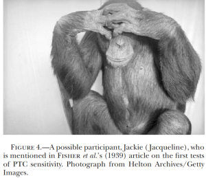
Be careful when you write the title to your scientific works (or any work) lest you produce something that can be misinterpreted like I misinterpreted this particular title, morphing it into this hysterical  concept of having a  “barbeque”.  After all it does say “taste-testing”.

So here is the title of the RA Fisher et al, article in Nature, way back in 1939. “Taste-testing the Anthropoid Apes.” R. A. FISHER, E. B. FORD & JULIAN HUXLEY . Nature volume 144, page 750 (28 October 1939).

What this silly interpretation led to was kind of interesting, and while I wasn’t about to pay 8$ to read the original Fisher et al article, I did find an open source article from 2003 (Stephen Woodling. Genetics 172(4):2015-23) that offered up some equally flavorful language from that 1939 publication. The old guys did not hesitate to “opine” in their publications and banter back in forth in actual publications.  The topic here was phenylthiocarbamine — which some individuals can taste as “bitter” and to others is completely “tasteless”.  These guys saw a pattern of inheritance, and one of the best known Mendelian (nearly simple) traits in humans, which paved the way for about 70 years of new investigations. Would love to have time and knowledge enough to read and interpret all these articles.

Human mitochondrial acetoacetyl-CoA thiolase

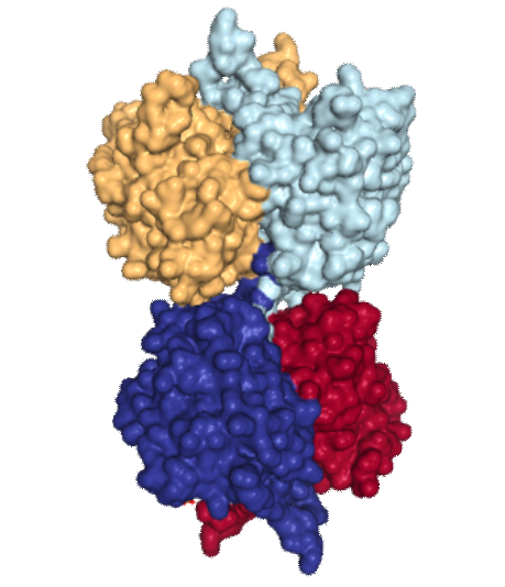
Human mitochondrial acetoacetyl-CoA thiolase: (found on the RCSB PDB – 2F2S)
This site is awesome, and I cant even begin to imagine how many smart people have worked tirelessly to make these data available to the average joe like myself.  Thanks to them.

The image of acetoacetyl-CoA thiolase of four different nearly identical chains identified by color in image to right.  I did not find articles (except one) stating that this was a membrane protein, and in which (outer or inner or cristae membranes) it would reside, and whether at the cristae pore or not.  My interest in posting pictures of this protein came after seeing that it has a 3dimensional symmetry that was visually quite wonderful.  Below are two sets of images taken at points of symmetry using the RCSB PDB link listed above. The symmetry is best seen with the ribbon molecules, but also is obvious with the space-filling and residue coloring modes.  I found this protein while looking for other mitochondrial membrane proteins, I am struck with how often nature mirrors flips and rotates basic protein elements to create function. In the images below molecules are colored as N-terminus (blue) to C-terminus (red); from 5′ end (blue) to 3′ end (red) — , bottom row is colored by residue. 3 green lines show symmetry (the line in z axis comes straight forward and looks like a point at the intersection of x and y. The green box shows a close vertical mirroring of the molecule…. rotated 90 degrees. I think the particularly interesting loops (four total) would have some special functions, seen at the top and bottom as presented in the ribbon diagrams.

Last image shows hydrophobic sites (right)… doesnt look like there is a region that would be membrane-bound does it?

It might sound silly to try to find out the size and shape of the known membrane proteins in mitochonria but just looking at the TEM images of mitochondria that I have accumulated over many years and many experimental conditions i am convinced that I can find some unique things to describe and show, but not knowing enough about the membrane proteins makes it less than satisfying. So at the beginning of the search, the ATP synthases was clearly fun, and easy to spot in some abnormal mitochondria.

Porn in advertising

There a few addvertisements and corporations that I feel really compelled to speak out agains. Victoria Secret, and now Amazon.  Victoria Secret’s adds I deplore, it is pornorgraphy, it also creates a need in women to objectify themselves, the very thing that some of us work very hard to prevent.  One add in particular made me vow never to buy anything from Victoria Secret…. when they used a christmas song to parade their naked women.  I did call their offices about that.

But Amazon…. why would they find it necessary to have a song  “you can ring my bell” that is just as pornographic as anything Victoria Secret puts in front of the camera.  I will voice my opinion, and never buy through Amazon.

Mitochondrial ribosomes: where was I

I googled “mitochondrial ribosomes electron microscopy” and expected the first citations up to be pretty recent…. nope, this one Kleinaw Neupert and Miller (not this miller) 1974 popped up.  Stating the obvious, muscles in the migratory locust certainly displayed many structures which were purported to be mitochondrial ribosomes.  Two particular images caught my attention: isolated cytoplasmic ribosomes and isolated mitochondrial ribosomes. In the “older” publications micron markers were required so this made it easy to determine the approximate insect cytoplasmic ribosome size vs that of the mitochondrial ribosomes (and at the same time look at the cristae for clues as to the ATP synthases-related bending and also to look at what might be something akin to mitochondrial pores at the base of cristae). Just as an aside here, the materials and methods sounded like a trip down memory lane for me, ha ha… though i used the Siemens Elmiskop model  1A introduced in about 1964, older than the 101.  Anyway some interesting measurements from an image here presented below.

About X=16nm is the distance between periods in the rhythm I see along side the mitochondrial RER tether.  This is based on the micron marker from the publication and also an estimate of ribosomal size from the micrograph itself  shown on the image.  Cytoplasmic ribosome about 21nm here, mitochondrial ribosome is about 15nm. Black bar micron markers 100nm and area enlarged is the box in the image at left. Left image has the mitochondrial portion outlined in red (as it is also in the insert with the cytoplasm there on the right side, mitochondrion on the left.