Monthly Archives: August 2018

Fuzzyballs and Superballs

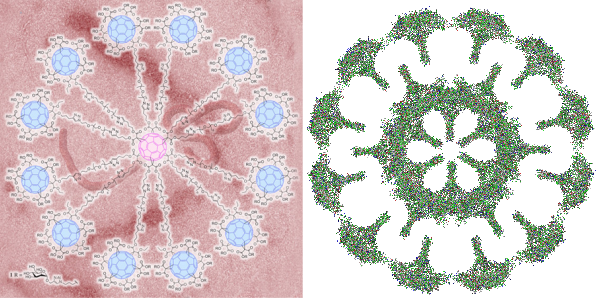
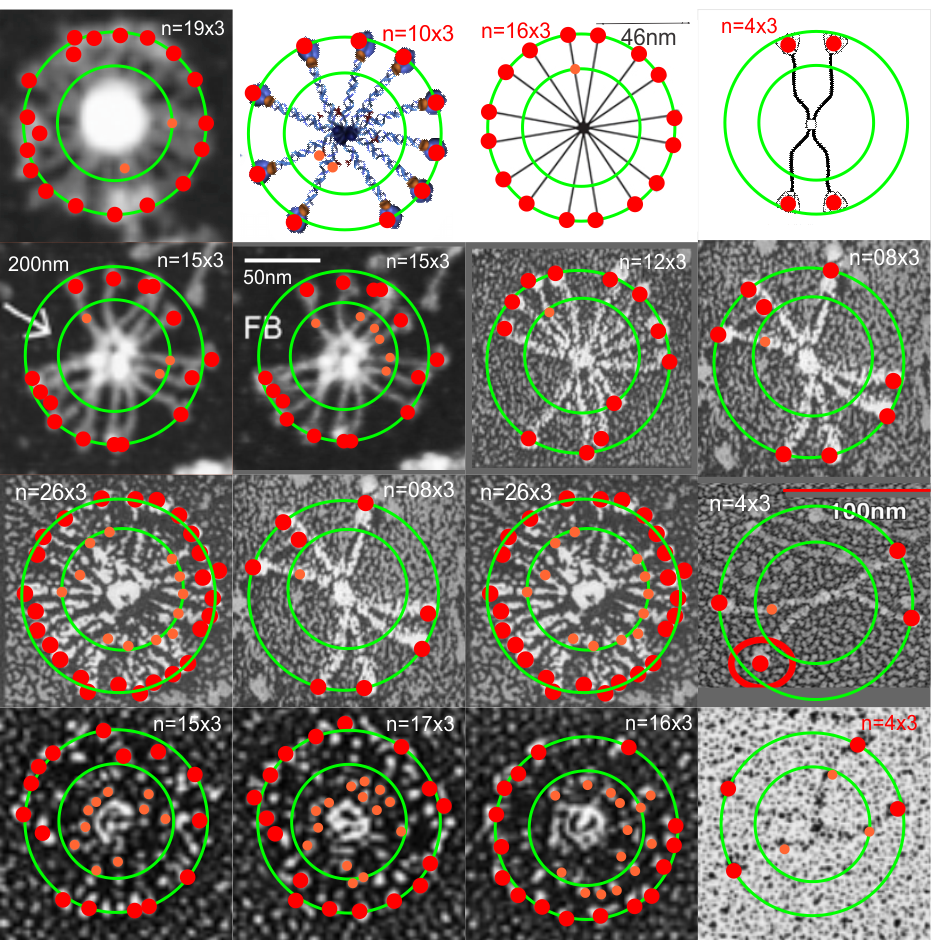
Two great types of structures, one nature-made the other man-made.  Here is a summary of one such nature-made structure, a fuzzyball, so called, which is a higher order oligomer of surfactant protein D.  One such fuzzyball also exists for surfactant protein A, a topic for another graphic next week.  So i scoured the literature to find a few diagrams and as many electron micrographs (usually shadow cast or negative staining) images of surfactant protein D as i could find to see whether there was a pattern to the assembly.

Almost the entire literature states that the oligomerization is from monomers>trimers> dodecamers>fuzzyballs (of different orders of oligomerization).  I have gathered such images and while the micron markers on all these different publications varies… consensus would estimate the dodecamer, as two trimers, mirrored, one carbohydrate recognition domain to the other, the N terminals in the middle to be about 100nm.  So i used this number (outer green circle in all=100nm – whether diagram or micrograph). In some images there was a little hint of something going on in the middle of the molecule so i marked these where see with an orange circle… and estimated with an inner concentric  circle where they might lie in the whole molecule.  Red dots are over the areas that would be the three different carbohydrate recognition domains (i should have used some three-part circle or similar to aid in the identification.. maybe i will swap this out).

The point here was to see whether there was a pattern (above the monomer, trimer, dodecamer, etc to oligomerization.  it seems that a fair statement would be 16 trimers…  I counted 15s and 17s, and 16s, so maybe the position of the fuzzyball as it fell to the 2D from its 3D made one or two difficult to differentiate. Of the three diagrams, the most right and the middle right have multiples of four….   the diagram to the center left looks like 10 to me, and probably 12 or 16 would have been more to the point…  I credit lots of authors with their images, none of which did i like to the original pdfs… as they were cut pasted, refined, cropped, adjusted for HSL, and features added….  so to me they are more than 10% changed. (LOL)..  The fuzziest fuzzyball gave me a count of 26 trimers…. but i bet is really more apt to be 24 or 28.  Two images show up twice, second and third rows down – just checking the various counts as repeats.  On occasion the original bar markers remain with the images.
Verge of a Dream: Taking one of the waves

I just hold the board
tightly, taking one of
the waves, without real
thought, parading toward
the shore. Maybe the sky
will darken tomorrow, maybe
I’ll take the car to some
point, to get a fresher
perspective, let the next
swell tell me what’s in
store. Then when the sun
decides to brigten, and
I feel I’ll turn to a
million pieces before I reach the
edge of the water running
hard with all that’s left
in me. With all that you
might contain but won’t.
Though you say hello
with a hug, once in a while.
A wet shoulder against
your cotton clothing goes
un-noticed the way it’s
night before or after
independence or christmas
day, I don’t remember
and in going by
it is also surprising
you stopped and told me so.

Babies hate broccoli – video

I was looking at this cute video with dozens of little kids who turned up their noses when being fed broccoli ..this was on  facebook (i don’t know if i could find it to post it here) but after viewing the video i began thinking about what would cause babies to reject a food that i personally think is a really healthful part of our diets. Truth told, i dont think my mom fed us much broccoli as kids, sadly, and i didn’t eat much of it until my seventh decade.

The babies universally made cute screwed-up expressions, when asked to taste broccoli and spit it out. I began to reason that this video send out some of that “fake news” that never gets straightened out, and the take home message which was “broccoli is horroble” was the “wrong” message.  The message here  “i hate broccoli” is not the message that should have accompanied the video.  Instead of “i hate broccoli” the message should have been something like this:

What a marvelous instinct has evolved in babies that protects them from their urges to put things unknown in their mouths when mom and dad are not watching.  Foreign objects feel, taste, and smell different than the bland diet they are used to and such odd tastes, shapes and textures  (particularly like broccoli), would trigger such a “warning” response that having a vigorous negative response to such items would indeed be a very good instinct to have.  I have read where exposure to new foods in infants requires an enormous number of “trials” maybe upwards of 17 — at least that is the number that comes to memory, before an infant or toddler (and some adults) will try to eat a new food.  A marvelous instinct that has undoubtedly saved many many children’s lives.

HERE IS A NICE ARTICLE ABOUT introducing new foods, and foods in general from TODAY’s PARENT but it doesnt address food rejection in infants. THIS ONE DOES.

My ancient mother Katrine (Katrina)

I am a descendant of Katrine (sometimes I find it is spelled Katrina) (K2a7). What an awesome time we live in…. to be able to find onesself in the vastness of humanity dating back to the stone age. Unvelievable, and thank you wikipedia.  I cant wait to add to this post. And a toast to my ancient grandmother Eve, or mt-Eve or  mt-MRCA, as the historical beginning of us all, again, thank you wikipedia.

K2a7 : found in France and Britain

Uncle Otzi : the iceman (“hypervariable segment (HVS-I). Two nucleotide transitions, at positions 16224 and 16311, indicate that Ötzi’s mtDNA belonged to haplogroup K, (but K1 apparently not K2 like me) a subclade of the major west Eurasian haplogroup U”)….

It would behoove us all to examine our expansive backgrounds and unity in ancestry, there are no walls, no borders, no exclusions… we are a collective humanity.  The dots on the map for my ancestors is enormous…. “I might be a lot like stone soup….. a little piece of this and that, from here and there, contributed by countless these and those”

“Haplogroup K originated in West Asia as a subclade of haplogroup U8b some time between 20,000 and 38,000 years ago……It is now certain that haplogroup K was a major maternal lineage of Neolithic farmers and herders before they entered Europe.”
This reference provides a long table of K origins, including this below which includes K2a7. (14,000 – 7,000 years ago; Behar, et al.)
K2

K2a : found in Late Neolithic England (Bell Beaker) and in Bronze Age Poland
K2a1 : found in Britain / found in EBA England
K2a2 : major Ashkenazi Jewish subclade
K2a3 : found in northern and eastern Europe
K2a4 : found in northern Europe
K2a5 : found in north-western Europe and Iran (Persians) / found in Bronze Age France and Russia (Fatyanovo culture)
K2a6 : found in the North Caucasus, central and north-western Europe
K2a7 : found in France and Britain
K2a8 : found in Spain and the Canaries
K2a9 : found in Neolithic Alsace
K2a10 : found in north-western Europe

Wikipedia states “A study involving Caucasian patients showed that individuals classified as haplogroup J or K demonstrated a significant decrease in risk of Parkinson’s disease versus individuals carrying the most common haplogroup, H”

L(mtDNA Eve)-N-R-U-K
Haplogroup K, formerly Haplogroup UK, is a human mitochondrial DNA (mtDNA) haplogroup. It is defined by the HVR1 mutations 16224C and 16311C. It is now known that K is a subclade of U8.

C reactive protein

Looked up to see whether this particular protein (a product of the liver) is elevated in lichen planus patients, and it was….. and while the linked article here states that this is correlated with an increase in cardiac rhythm…. i bet it is not the disease itself but the inflammation in general… as they say this is also true for other diseases.  So i have to agree, this is a stressful disease….  the association shown here in a small study is probably a “whole body” response to having an immune disease, maybe any disease, or in other acute inflammatory states.

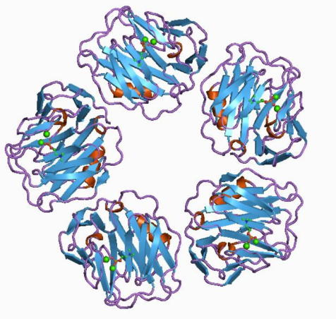
wikipedia says: C-reactive protein (CRP) is an annular (ring-shaped), pentameric protein found in blood plasma, whose levels rise in response to inflammation. It is an acute-phase protein of hepatic origin that increases following interleukin-6 secretion by macrophages and T cells. Its physiological role is to bind to lysophosphatidylcholine expressed on the surface of dead or dying cells (and some types of bacteria) in order to activate the complement system via C1q.

CRP is synthesized by the liver in response to factors released by macrophages and fat cells (adipocytes). It is a member of the pentraxin family of proteins. It is not related to C-peptide (insulin) or protein C (blood coagulation). C-reactive protein was the first pattern recognition receptor (PRR) to be identified.

The protein itself is beautiful…. i think i might just honor it by constructing a stained glass rose window pattern.  ha ha.  thank you wikipedia for the image.

Lookie there, my cover inside the July 2018 edition of Microscopy Today

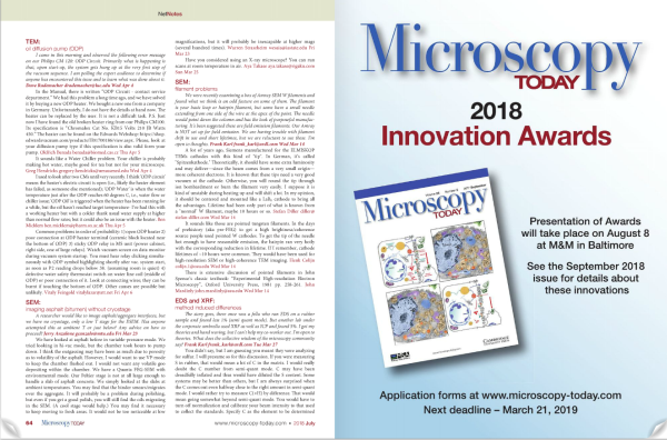
“Lookie there, my cover inside the July 2018 edition of Microscopy Today”

I was looking up the editorial staff of Microscopy Today to see if they would be interested in considering a short editorial (which i would love to name Ecc 1:9, but will probably not use that for reasons of pseudo “separation of church and science” LOL).  And filling through the online pages of the July 2018 issue i ran onto my own cover on one of the inside pages.  Awesome.   I made a link to that issue, but i don’t know if it will be held in perpetuity or whether it will rotate with new issues.

Molecular and macromolecular architecture follows patterns

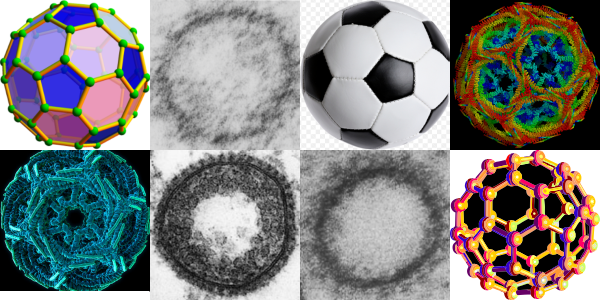
This is a wild goose chase looking for structures which explain and identify those which I have seen repeatedly in hepatocytes (and other cells), this time including clathrin coated vesicles and COPI and COPII vesicles.  I am continually amazed how certain motifs repeat themselves in the microscopic, molecular and modern world.  Stability in structure seems to transcend size.  So image below is a collection of fullerene diagrams, some from my own electron micrographs, one from a widely republished electron micrograph (credits to the author if i knew who it was) and some from various wikipedia posts and of course the most widely recognized fullerene structure… the soccer ball.   Just for fun but also tying in the concept that nature made it first. There is a difference in magnification from the upper left (i think around 1 nm – which actually sounds too big to me) — to the soccer ball 22 cm in diameter — well you can do the math  2,200,000,000 nm.

Superballs vs fuzzy balls: immunity

I got into searching for the structures of clathrin, COPI and COPII and one website visit after the other led me to this link. There headline was “Globular glycofullerene molecules prevent virus from evading immune system and entering cells” and I got goosebumps when i thought to myself that the surfactant protein A and surfactant protein D fuzzy balls might very well act as these proposed superballs in inhibiting infections from those viruses which have developed the reverse method for “entering” cells and avoiding the immune response (ebola and HIV for example).  Seems a really fun thing to research.  I posted a picture of what i designed as a surfactant protein A fuzzy ball (surfactant protein D makes fuzzy balls as well) and immediately saw the similarities between the octadecamer glycoprotein protruding  in a sphere in both these fuzzy ball naturally occurring innate immune functioning proteins and the one that was produced by synthetically.  Nature figure it out first.

BTW i dont like how the EBOLA virus is tangled within the backdrop of the globular glycofullerene on the left, called a  “superball”,  in fact it is kind of “nonesense” to me…. I could however envision the EBOLA virus stretched to oblivion as if stuck to a round velcro sphere, with areas exposed for digestion and removal.  It has been stated that EBOLA type viruses (EBOLA is a “Filoviridae” virus and older than previously thought). That group of viruses has been interacting with mammals for several between 5 – 23 million years and it makes perfect sense that some sort of defense mechanisms have arisen in parallel. In fact a little bit of searching shows up this reference on filovirus entry.

s