All these snowflakes used in a diagram (interlaced hexagons) were taken by a professor at Cal Tech. I contacted him for permission to use his “real” images. No monetary gain for me was derived, but I thoroughly enjoyed using the variety that nature has to offer and that he captured on camera and posted online. Also thanks to Mike Neer who is editor of this CorelDRAW help magazine, as he let me play around with images for a few of the covers.
Daily Archives: September 25, 2018
Click 5 – aka casper the friendly ghost
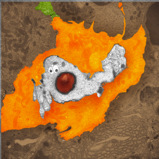
One fun image from long ago, never a cover illustration, but could have been one at some point, particularly for a fall issue of a throw-away-journal. This is actually from a micrograph taken from a KO mouse CLICK5. The particulars of that particular KO mouse have long passed from my memory, but constructing the image is still fresh. ha ha..
White part is the parietal cell nucleus, nose is red nucleolus) cytoplasm is pumpkin orange, and the green stem is a portion of of another cell.
Skin cancer clock – illustration for cover
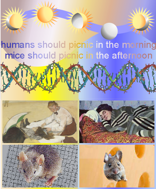
Not all illustrations make it to the journal covers. Going back half a dozen years I found this illustration (requested by Aziz Sankar) about biological clocks, UV exposure and cancer. My illustration did not make it onto the cover of proposed journal (at this point I cannot even remember which journal it was submitted to) but when I saw it i thought… what a cute attempt. So here it is anyway.
Off-center N terminals of SP-D fuzzyballs
 There is nothing I am doing here but questioning the current notion of how surfactant protein D is arranged into fuzzyballs. I have none of my own images to present, just those published by others which count is close to 75, from several authors.
The publication of Arroyo et al that I have mentioned many times has very lovely AFM micrographs, and they have provided counts of 600+ SP-D images, and it is from these the few of these they published that I am making suggestions that perhaps the N terminals of surfactant protein D molecules are NOT dead center, but arranged in a sort of “ring” near the center instead. The other suggestion is that the arms of the surfactant protein D hexamers (maybe dodecamers) are not straight rods, as almost universally depicted in diagrams of SP-D, but rather are gently curved, almost as if they are hexamers whose interaction at the center is tangent to a center sphere, but at an angle of 60-100 degrees. In measuring the angles in just this one fuzzyball, the average degree was about 67o. (lower left is measured from the dotted lines in upper left — nb, this was just one fuzzyball, and there are many ways to draw those arcs than the way I drew them the first time.)
Image on the left (with its dotted lines) is almost identical to the one posted before (here) but i changed the bending of some of the dotted lines to what I thought was a better fit. In addition I added two more images, with different contrast, and outlined in blue the areas where the N terminal portions of the hexamers make a tangential connection with each other. I think that this (and other fuzzyballs I have observed) pretty much show up as multiples of 2 which is what might be hexamers. Of those which I have currently measured, there will be an angle between SP-D arms, but I will also measure them again, thinking of the molecule itself as an entity which is made up of slightly curved hexamers (or possibly dodecamers), and the angles will certainly be different.
One thing that makes the N terminal attachments NOT being dead center is the presence of slightly brighter areas near the N terminals, which could be representative of the glycosylation sites. For small bright areas to show up at a distance which is proportionate to the ring-center, is encouraging.
Lower right figure has a single hexamer (bent) in which i have measured the angle, and also outlined the bright areas of the N terminal(s), and possibly the glycosylation site(s).
I am impressed by the fact that the CRDs are “lumpy” in this particular image, and I have traced around them, there are almost suggestive of the three CRD domains of the trimer.
One thing that is easy to question on this post is why i drew the arcs with a convex curve and why the sample of a hexamer looks like the curves are concave..haha good question.


