Daily Archives: July 18, 2019

Arm length of SP-D, conglutinin, CL-43

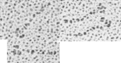
Holmskov et al published images of CL-43 and SP-D and conglutinin prepared in hopefully all in the same manner of rotary shadowing technique and magnification, and i took each of those trimeric arms measuring them separately (using a line with sufficient numbers of nodes to account for the curvature of the molecules, from the edge of the CRD to the center (presumably the N terminus).  The arms of SP-D dodecamer were each measured and a mean found, similarly for conglutinin, and 4 individual arms of CL-43 taken at random from his picture (60nm, 47nm and 46nm respectively).  The micron marker is hopefully accurate, and it does then show a difference in arm length with each trimer of SP-D at about 60nm (making the dodecamer about 120nm in diameter) the conglutinin arms individually at 47nm, the full diameter at 91nmn, and the CL-43 very close to the conglutinin at 46nm (no dodecamer found in his image). (you can see his figure numbers still on the unretouched images).

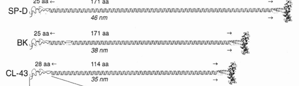
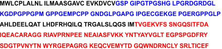
So a really easy way to measure the length of a curved c-type lectin arm (including bends and kinks) is to create a vector line with nodes at places which accommodate the shape (this is a different method than calculating the arc angle and arc length that I tried previously. This is quite easy by comparison. This group published a diagram with relative sizes here  as well which also has micrographs accompanying, maybe some of the same images – i will check and measure. I know at one point i did not agree with the length of the collagen-like region, and had difficulty matching that with the AFM images produced by others.  His diagram of the AA sequence size comparison i have posted before, but include at the bottom.  THey are pretty close to what i measured. Relative to their SP-D being a single trimer of 60nm in adjusted length (adjusted for curvature) then the CL-43 is 46nm as a single trimer – exactly what his diagram would suggests (if SP-D is 60nm, then CL-43  is 47nm),

Images below


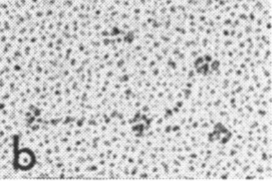
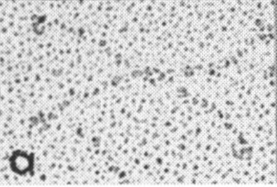
From Hansen and Holmskov (Immunobiology, 199, 1998)

Astounding inaccuracy in a scientific diagram of surfactant protein A

Remember playing “telephone” as a kid (also called “chinese whispers” — the title of which in and of itself denotes confusion)? Here is a link to wikihow if you never played it. The purpose is to show “in real life” what happens when information is casually passed from one to another (as in gossip… and i am calling the remake of scientific illustrations from previous publications as a similar phenomenon).  The important truth is that information is easily corrupted. No more description is needed for the image below which is supposed to portray surfactant protein A.  Clearly this artist/scientist team knew very little about SP-A or they were playing “telephone” with the specifics of how to draw the diagram.

What is sad to me is that this really bad diagram shows up in a place where many individuals might actually expect accuracy (a site called Global Science Books ) ? A link to the pdf that I found is HERE.

I will list what I think they missed, data from SFTPA1 online and the literature.

  1.  The ratio of AA for each of the 4 domains seems to be wrong, as the N terminus is very short, the collagen like portion is pretty long (blue), coiled coil neck domain (black) is not too long, the CRD (red) is quite long.  The relative distances of these four domains in this diagram are quite far off, in spite of the fact that many much better diagrams are available for free. Signal peptide is 1-20.
  2. Where did the third strand of the collagen-like domain go? It looks to me like the two twined black lines in their diagram are just that “2” not “3” as repeatedly mentioned in texts (every text i have read in fact) and why would it be called a trimer if not three, and how in the world does the third CRD domain hook up when there is no line in the collagen like domain to which to attach it?
  3. There is virtually no distinction between the winding of the collagen-like domain and the coiled coil neck… an arrow points to the transition, but there is not definition, and as well, there are only 2 of the 3 strands in the neck domains present.
  4. It is difficult to interpret the trunk like portion of this diagram, and how it might relate to the interactions between the collagen like domains, of the 6 trimers. It might be noted that in their diagram (not shown here) lying right next to this SP-A diagram shows the dimer of SP-B in the same two lines as the trimer of SP-A? Why?
  5. What in the world are the “balls” circles at the N terminus? Is this a depiction of N termini binding sites?
  6. Why are the three supposedly nearly (if not absolutely) identical CRD pictured in different sizes?. If it is an attempt at forshortening in graphic design if fails miserably.

OK, this is enough, you get the point.