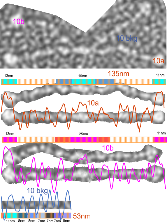
LUT tables are collected from AFM images, and provide detail that is interesting and likely informative. I wondered the same about shadowed TEMs. Here is clue as to whether any information about SP-D dodecamers (or other multimers or trimers or hexamers) can be derived from shadowed images. It is pretty clear that a background LUT plot of the same micrograph in the same area (blue lines) does have a repeating pattern of the deposited shadowing material. The Ntermini that meet in the middle of the dodecamer and the CRD (of which there are 4) do show a different dimension than the regular repeating 8-10nm background. How well the shadowed images predict the position of the presumed peaks between the Ntermini and the CRD could probably be worked out.
These particular images are derived from a micrograph published by E. Crouch et al, so further LUT analyses might show something about the full structure of SP-D which is not yet available on RCSB. (i am not sure why since it is pretty clear that the collagen-like domain presents at a slighly straight to curved portion of the dodecamer. What also might happen is that the Ntermini might bind side to side, either in addition to or instead of end to end.