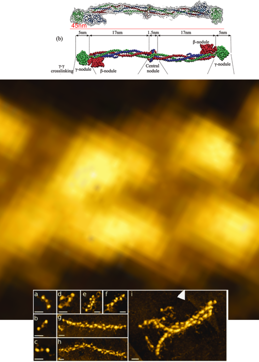
Atomic force microscopy images of fibrinogen, fibrin oligomers, and protofibrils and reconstruction of a protofibril model. (a-i). Images by high-resolution atomic force microscopy (used from a second hand image posted from original of Drs. Anna D. Protopopova, Nikolay Barinov, Dmitry Klinov) from a post on Research Gate.
Sometimes putting the molecular model with the AFM image is a little difficult. Using the measure of 45 nm for the molecular model at the same magnification as the micrograph is marked (bar=30nm) something just looks out of proportion, however, the length of what would show up as between-peak brightness probably does equate to the model, but what is puzzling is the thickness of the two strands… model vs micrograph where just a tiny portion of the micrograph (about the middle of the original (shown below) was cropped and enlarged to scale of the molecular model diagrams.