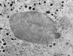
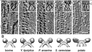
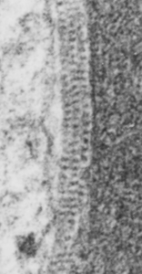
Mitochondria from vinyl chloride exposed animals have been posted on this blog before. HERE and HERE. I have made the assumption (no proof what-so-ever) that this just seems a perfect match for ATPsynthase. Here is another example of such a mitochondrion, also showing the highly organized repeating pattern found in the intra-cristal-space of mitochondria from other liver electron micrographs from guinea pigs treated with vinyl chloride. Amazing similarity. THis waits for someone else to confirm, but in the meantime when I see the images, I will post. Someone out there will know. So here is a mitochondrion with such a cristi, and below that is someone elses micrograph (publication site linked) which shows the spiral repeating structure of ATPsynthase.

REF
 One of their figures (not pictured here) shows about 16 dimers per 200 nm = 1 per 12.5 nm. When I measur
 ed the ribosome at 27 nm in my image (to the left) of intra-cristi structures, then there were about 19 densities per 200 nm. So the numbers don’t add up too well. Particularly with one of their figures copied above where their measurement (white bar in the figures and insets above) is noted in their publication to be 50 nm. This is quite far off what I see in the intra-cristi space in the figure to the left. It also seems clear that they have a propensity to be found in these groups between the outer and inner mitochondrial membranes and less frequently on the inner cristi.