Category Archives: Ultimate order, the cell

The beauty and order of life is astounding.

Surfactant protein D – fuzzyballs

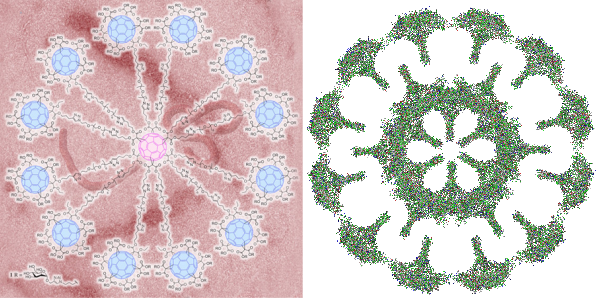
Arroyo et al have published what I think is a great paper using atomic force microscopy to look at many different multimers of surfactant protein D.  There are a couple of things that I think they saw that might be what I also noticed looking at so many images of SP-D with various EM methods.

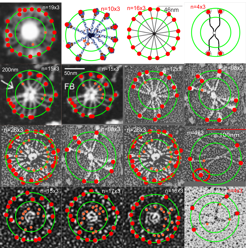
That is, there seems to be a blip or bulge in the SP-D structure close to the N terminal, and I marked that with some images in a previous post, then removed them for a summary page and the early video clip of SP-D.  I think they are real, and also this publication indicates that it is the N glycosylation site…. and used some experimental manipulations to justify that view. Another issue is that the N-terminal sites of SP-D dont really often line up into a single area, they look sometimes like they are groups of separate arcs which have an angle of about 40 degrees (as measured on the micrograph below (the small angles) two angles were about 120 degrees.  The center doesn’t look like a concentric area, rather a radially spaced loop-association.

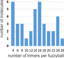
This micrograph is from Arroyo et al, and it shows (as do many other images from their publication) what I am trying to indicate. Actually many of their fuzzyball micrographs show this the inner segmented ring rather than a single central density. I used their picture,  and photoshopped it to eliminate background notations and debris, and other nearby molecules (you can see these in their original picture so you can compare) but I did not change the image of the molecule in any way.  SO: one of the big issues is the curviness to the arms of the SP-D molecule (see my blue line in the image below), and another is the-off-center radially arranged N-terminal densities. These just make it unlikely that there is a “center” point where the N terminals come together in a single lump space.  A third issue is that it seems like the angles between the hexamers (if in fact that is how they assemble into fuzzyballs… one hexamer at a time, or perhaps one dodecamer at a time, where the the angle is not that “acute” but is pretty broad, maybe on the order of 40 degrees.  This is what the dodecamer shows most typically so there may be a reason to assume that in the fuzzyball that the 40 degree angle for the four arms (and a broader angle of 140 degrees between arms) is maintained. Blue ring (that I have added to image below) might be where other interactions in the fuzzyball structure bring the structure together in a sphere? or disc? at points of intersections of the “loops”.  Other loops and centers around the blue ring are shown with white dotted lines.  This particular fuzzyball one would prefer to see with even numbers of arms (they may be there)…. but apparently an arm or a couple of them got misplaced in the preparation process (LOL)… it is also my suspicion that there is a quantity  “four” –  that is, that the elements are divided into quadrants as reasonably convincingly shown in the micrograph below, and that works out to the 16 arms (an even number and multiple of 4) which pervades the surfactant protein D images that I have collected so far. White bar is 60nm according to Arroyo et al’s original image.  Blue circle is about 40nm relative to their white bar marker.

organization of arms of surfactant protein D fuzzy balls electron microscopy


Are there angle restrictions on distribution of SP-D trimers in fuzzyballs

Looking through as many SP-D images as I could find in the literature I began to sense that the distribution (radial) of SP-D dimers was not random. I am determining the angle of separation of each arm of a SP-D fuzzyball to see if the numbers fall into classes of small, and larger. The data for these three pictures is spot-on.  Various authors are responsible for these electron micrographs, i give them credit..they are not my personal photos. Images on the right and left are shadowed, image in the middle is AFM.

To create the round “generalizations” of angles in SP-D fuzzyballs, the actual angles were measured in the molecules below.  A mean was obtained for each angle-set: e.g. the center pix had 4 measurements, as follows:  I will measure all 90 images and see if the trend holds.
147.46
127.6
41.62
43.32

Sometimes I laugh – SP-D

I am trying to write a comment page about innate immunity proteins and the possibility of nano-superballs being used to configure and distribute such immune proteins (which, by the way is a logical next step in that research since at least two of the C-type lectins that are important for innate immunity actually do have fuzzyball multimer structures. Those would be SP-D and SP-A. When I saw this cute dimer of surfactant protein D I just laughed to myself, such a happy dancer.  This particular image of a surfactant protein D dimer came from a portion of an AFM image published by Arroyo et al, JMB, 430:1495-1509, 2018. Thanks to them for the laugh, and the inspiration to keep up the exercise program for the sake of good health.  This guy (girl) is just about to cross the half-marathon finish line.

What is interesting is the light and airy feeling, arms outstretched, legs in the air and the perfectly positioned ball (likely a loose CRD, ha ha) for a head.

Surfactant protein D fuzzyballs

I have scoured the literature to find any images, and even diagrams, of rat native and recombinant surfactant protein D, hereafter called SP-D. There are some very nice images (perhaps the best) by Crouch et al, published in JBC, (also using recombinant SP-D) which I have cut and pasted into this video, counting all the trimeric arms with “attached” carbohydrate recognition domains. Crouch et al indicated that about 5% of the structures are multi-assemblies, that is something around 12 arms? or more is what they show. Actually two authors have commented on the fuzzyball rarity in shadowed images for microscopy. Other images came from google searches for SP-D diagrams, and many of these are from McCormack, and from others in the lab here at Cincinnati’s Childrens Hospital.

The three dimensional structure (multimers) of SP-A and SP-D have been shown to be important for function.  Vieira et al 2017“In conjunction, these data demonstrate the critical role of oligomeric  and tridimensional structure of surfactant protein A and surfactant protein D for proper function.” and also say “SP-A and D can target pathogens by simple aggregation, without a direct interaction with the cell. Similar to complement C1q, SP- A can function as an “activation-ligand” which facilitates particle uptake once it is coated by Immunoglobulin G”

SP-A and SP-D have distinct functions though both are quite easily given the “title” of multivalent innate immune proteins.  What is interesting is that under some circumstances and not others, they may adopt a more spherical  and highly oligomerized state, or they may remain trimers, hexadecamers, dodecamers and in lower oligomerization states. For some reason the bouquet for SP-A as an 18-mer is most frequently cited in the literature when in fact the electron microscopy might show full fuzzyballs of SP-A, and other forms (periodicity in flat sheets, under other circumstances.  In the same micrographs of type II alveolar cells, SP-D fuzzyballs really havn’t been encountered, but in vitro SP-D makes great fuzzyballs.


https://thankuscience.com/wp-content/uploads/2018/09/SP-D.avi (cut and paste)  SP-D .avi link

I was curious whether there was a pattern to the number of trimers in any given fuzzyball and for SP-D there were numerous articles which actually had pictures of oligomers.  The number of arms (trimers) for each fuzzyball of SP-D, randomly encountered in publications (not to be confused with randomly photographed under the microscope… as i am quite sure there was selection bias in photographing these) is shown on x axis of this quick summary graph.  Incidence is on the Y axis. I could have counted more (and may do that) in some images where there was indication that perhaps there were arms and CRDs that had been displaced during collapse of the fuzzyball onto the grid. Image below is taken from Crouch et al mentioned above and used without permission.

Here is a short videoclip of SP-D fuzzyballs — you can see where I have counted the carbohydrate recognition domains, as I overlayed them on the existing micrograph with red dots. There are places where I might add or subtract one now and then from what is pictured, but in general you can see that top number is something around 26 arms (each a trimeric molecule) in some fuzzyballs. I think more than 32 (a multiple of 4 is likely) arms in any given fuzzyball is going to be pretty uncommon.  It is very apparent in some that groups of 4 arms persist.  While the orientation of SP-D arms is probably end to end in those groups of 4, I am thinking that SP-A fuzzyballs are going to be four groups of 18-mers. Whether such is found in the literature I will soon find out.

Fuzzyballs and Superballs

Two great types of structures, one nature-made the other man-made.  Here is a summary of one such nature-made structure, a fuzzyball, so called, which is a higher order oligomer of surfactant protein D.  One such fuzzyball also exists for surfactant protein A, a topic for another graphic next week.  So i scoured the literature to find a few diagrams and as many electron micrographs (usually shadow cast or negative staining) images of surfactant protein D as i could find to see whether there was a pattern to the assembly.

Almost the entire literature states that the oligomerization is from monomers>trimers> dodecamers>fuzzyballs (of different orders of oligomerization).  I have gathered such images and while the micron markers on all these different publications varies… consensus would estimate the dodecamer, as two trimers, mirrored, one carbohydrate recognition domain to the other, the N terminals in the middle to be about 100nm.  So i used this number (outer green circle in all=100nm – whether diagram or micrograph). In some images there was a little hint of something going on in the middle of the molecule so i marked these where see with an orange circle… and estimated with an inner concentric  circle where they might lie in the whole molecule.  Red dots are over the areas that would be the three different carbohydrate recognition domains (i should have used some three-part circle or similar to aid in the identification.. maybe i will swap this out).

The point here was to see whether there was a pattern (above the monomer, trimer, dodecamer, etc to oligomerization.  it seems that a fair statement would be 16 trimers…  I counted 15s and 17s, and 16s, so maybe the position of the fuzzyball as it fell to the 2D from its 3D made one or two difficult to differentiate. Of the three diagrams, the most right and the middle right have multiples of four….   the diagram to the center left looks like 10 to me, and probably 12 or 16 would have been more to the point…  I credit lots of authors with their images, none of which did i like to the original pdfs… as they were cut pasted, refined, cropped, adjusted for HSL, and features added….  so to me they are more than 10% changed. (LOL)..  The fuzziest fuzzyball gave me a count of 26 trimers…. but i bet is really more apt to be 24 or 28.  Two images show up twice, second and third rows down – just checking the various counts as repeats.  On occasion the original bar markers remain with the images.

Babies hate broccoli – video

I was looking at this cute video with dozens of little kids who turned up their noses when being fed broccoli ..this was on  facebook (i don’t know if i could find it to post it here) but after viewing the video i began thinking about what would cause babies to reject a food that i personally think is a really healthful part of our diets. Truth told, i dont think my mom fed us much broccoli as kids, sadly, and i didn’t eat much of it until my seventh decade.

The babies universally made cute screwed-up expressions, when asked to taste broccoli and spit it out. I began to reason that this video send out some of that “fake news” that never gets straightened out, and the take home message which was “broccoli is horroble” was the “wrong” message.  The message here  “i hate broccoli” is not the message that should have accompanied the video.  Instead of “i hate broccoli” the message should have been something like this:

What a marvelous instinct has evolved in babies that protects them from their urges to put things unknown in their mouths when mom and dad are not watching.  Foreign objects feel, taste, and smell different than the bland diet they are used to and such odd tastes, shapes and textures  (particularly like broccoli), would trigger such a “warning” response that having a vigorous negative response to such items would indeed be a very good instinct to have.  I have read where exposure to new foods in infants requires an enormous number of “trials” maybe upwards of 17 — at least that is the number that comes to memory, before an infant or toddler (and some adults) will try to eat a new food.  A marvelous instinct that has undoubtedly saved many many children’s lives.

HERE IS A NICE ARTICLE ABOUT introducing new foods, and foods in general from TODAY’s PARENT but it doesnt address food rejection in infants. THIS ONE DOES.

Superballs vs fuzzy balls: immunity

I got into searching for the structures of clathrin, COPI and COPII and one website visit after the other led me to this link. There headline was “Globular glycofullerene molecules prevent virus from evading immune system and entering cells” and I got goosebumps when i thought to myself that the surfactant protein A and surfactant protein D fuzzy balls might very well act as these proposed superballs in inhibiting infections from those viruses which have developed the reverse method for “entering” cells and avoiding the immune response (ebola and HIV for example).  Seems a really fun thing to research.  I posted a picture of what i designed as a surfactant protein A fuzzy ball (surfactant protein D makes fuzzy balls as well) and immediately saw the similarities between the octadecamer glycoprotein protruding  in a sphere in both these fuzzy ball naturally occurring innate immune functioning proteins and the one that was produced by synthetically.  Nature figure it out first.

BTW i dont like how the EBOLA virus is tangled within the backdrop of the globular glycofullerene on the left, called a  “superball”,  in fact it is kind of “nonesense” to me…. I could however envision the EBOLA virus stretched to oblivion as if stuck to a round velcro sphere, with areas exposed for digestion and removal.  It has been stated that EBOLA type viruses (EBOLA is a “Filoviridae” virus and older than previously thought). That group of viruses has been interacting with mammals for several between 5 – 23 million years and it makes perfect sense that some sort of defense mechanisms have arisen in parallel. In fact a little bit of searching shows up this reference on filovirus entry.

s


Certifiable? or does rRNA on RER rock

I was looking at liver electron micrographs, looking for mitochondria, trying to see whether cristae juntions are such a noticeable feature as is claimed, boy I can not find them easily. But in “boredom” not really boredom, more frustration, I decided to punch in a “kick” beat to the spacing of ribosomes along the RER membrane.  I hope this fun time with Fruity Loops doesn’t distract me from doing real science…. as I can see how fun it would be to use a top hat, steel drums, cymbals, for some ribosomes a little further away, and a base beat for those that are very clear and a blip for those that are faded out and then play two tracks together (opposite sides of the ER membrane being a separate track each). Now you know why I chose the title for this post “certifiable” (i am not crazy, as sheldon cooper says, my mother had me tested),

Mitochondrial interactions with RER

Hepatocyte here, GCLC ko mouse, shows the donut and irregular configuration that is common in circumstances where mitochondria are stressed (I personally have seen it several times but in unrelated experimental circumstances so it is probably a generic response mostly).  In this really opportune section one can see a mitochondrion (with an odd donut shape) ont eh left, and a section of RER which has been sectioned tangentially showing the closeness and absolute regularity of the ribosomes along a spiral of mRNA.

I bet at some point all information about these association and the proteins (i saw a list that mentioned in the 800s and counting) in the inner and outer and cristae membranes, and the cristae junctions as well as the matrix will be modeled.  Until then, circumstantial evidence for the power of the interactions between mitochondria and other organelles (and cytoskeletal proteins) has to suffice.

The ribosome spirals here look to have approximately 7.3+/- .47 (SEM) ribosomes per spiral, n=9 (a small sample but the best orientation, and a single micrograph…. so this is just a suggested number obviously.  19735_73218_#201 liver alb+/- Gckc -/- postnatal day 28. liver mouse, no NAC.

One thing abou these mice that is pneumonic is the dilated ER, a mix of smooth and rough, ribosomes spaced and the presence of the little bubble-blip invaginations of ER within the outer RER membranes.  These mitochondria also display fission lines and tubular cristae, and quite a bit of it.  Blue dotted line (outline of one part of a mitochondrion – that one tangentially sectioned beside the RER, white box, area for enlargement to the right. Upper image on right has cytoplasmic ribosomes in mRNA-spirals (orange) and lower image is contrast enhanced to highlight the spirals of cytoplasmic ribosomes.  I really don’t think there are any good examples of mitoribosomes in either of the two mitochondria shown here.

Numerous proteins in the outer mitochondrial membrane (encoded by nuclear DNA) target and or are attached to cytoplasmic ribosomes. Cytoplasmic ribosomes have been visualized on mitochondria membranes (that would be the cytoplasmic face of the OMM). These are suggested to be linked by the translocase of the (outer) mitochondrial membrane (TOM) and it is reported that the ribosomes are in clusters (Till Klecker et al, 2014 Trends in Cell Biology) and that pretty much looks like these nRNA-ribosomal spirals… tidily wound.

MDVs, mitochondrially derived vesicles (about 70-150nm) are  purposeful buds of membrane derived from mitochondria which are targeted to lysosomes, and maybe other organelles.

Just an “aside” here, but is it possible that the donut shaped mitochondria (invaginations, and extra turns and indents) might be somehow a deliberate attempt to increase surface area for interaction with other membranes.   Another question is the orientation of cristae.  I have looked for cristae pores, and that relationship…  just not seeing it overtly.