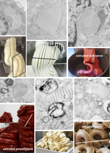
A summary and diagram – two years in the making (not so LOL)
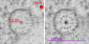
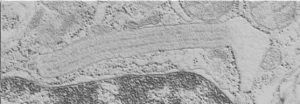
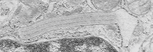
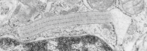
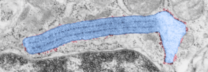
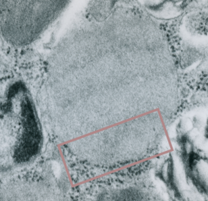
A protein granule exists in the rough endoplasmic reticulum of alveolar type II cells of some species which has remained relatively obscure. It has characteristics of a highly self-associated protein with a predictable repeating substructure and displays at least 8 prominent patterns:
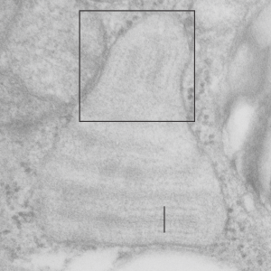
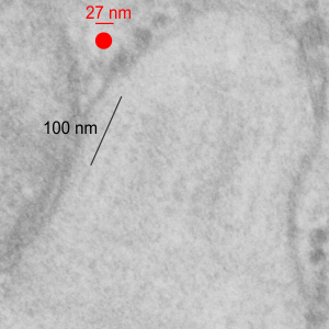
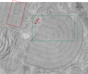
1) most commonly it appears as a layered sheet about 100 nm thick
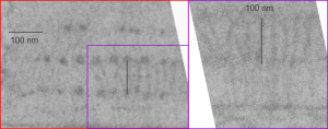
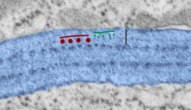
2) which upon cross section comprises two dense outer layers, 100 nm apart, a less dense central layer which can have up to 3 faint layers on either side. This pattern forms one period
3) the layered sheet (100 nm-thick period) can also be stacked many times creating a granule with a length and height of many microns
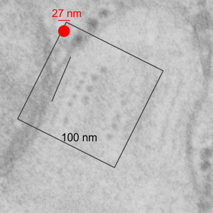
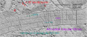
4) whether single or highly stacked, ribosomes only lie on growing ends of the granule and in opportune cuts a single period has 4 ribosomes, 1 at each dense layer and one on either side of the central layer
5) the perimeter of the granule could be linear, or rounded, with sheets (layers) arranged in patterns reminiscent of flow extrusion, where each of the 4 ribosomes equates to a die, and as if the rate of protein translation/modification, and the shape, size and press capacity of the ER determined if the granule would be gently curved or U shape, looped or folded-back, concentric, or branched. Some granules were a mixture of all these forms, even rarely intersecting
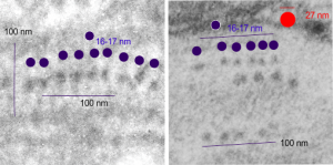
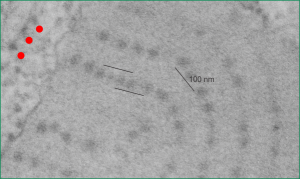
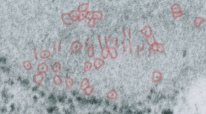
6) Both the outer dense, and less dense central layers appeared to be continuous on perpendicular cuts, but clearly punctate when cut tangentially
7) Outer dense bands showed a periodicity of about 4 per 100 nm, while less dense central bands showed a periodicity of about 5-7 per 100 nm, possibly lined up in a staggered format
8) A faint vertical tie was also seen at about 4 per 100 nm beginning typically at one of the densities
Measurements have been made but these are approximations derived from hundreds of micrographs and numerous publications. Similarly, measurements by numerous reports on the size and structure of surfactant protein A have shown that it is likely to be an 18-mer with a spread of the CRD at the top of the bouquet of something on the order of 25 nm. It doesn’t take a big leap to put four 18-mers together vertically and copy them side to side to come out with a pretty awesome banding pattern that fits the 100 nm dimensions.

