Category Archives: Science and art cover illustrations

…and yet another cover submission for the manuscript on SP-A granule and electron microscopy

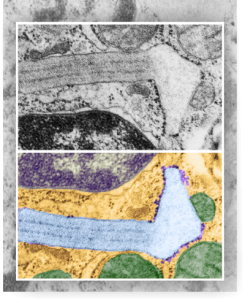
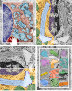
…and yet another cover submission for the manuscript on SP-A granule and electron microscopy.  This one is a little more dramatic than yesterdays post from the bottom right part of the image.  I like it better. It shows the quintessential granule, in black and white at the top, color at the bottom in a vertically mirrored image, directional ribosome attachment, electron-lucent areas under the non-oligomerized protein part, distinct periodicity to the dense bands (three complete “periods” in the lengthwise portion of this granule) and also the periodicity to the dense layers (3-4 dots/100nm) and even some of the faint periodicity found in the central lighter layers. In this electron micrograph one also sees part of the nucleus (bottom), a couple of nuclear pores, and some perinuclear space (but no granule formation in the perinuclear space is seen in this image, but it is found there frequently) and on the top, portions of three mitochondria and one adjacent to the lucent area of the granule. There are also other profiles of RER, coming and going which at some point might connect up to layered putative SP-A protein granule structures.

Four cover submissions for the SP-A manuscript: love the colored electron micrographs of granules

Four cover submissions for the SP-A manuscript: love the colored electron micrographs of granules. I am submitting this to Anatomical Record. They request a cover submission at the same time as initial manuscript submission, apparently, at least those are the instructions.  Typically I think the manuscript has to be accepted before cover submissions are made.  Oh well, I am just hoping for the best.  Preview here.

Electron micrographs of golgi and golgi stress

Cover submission from the early 2000s for a published manuscript by Shull Miller and Prasad where golgi, golgi ion transport, and golgi membranes (yep, i am not capitalizing the word Golgi any more, with all due respect to Dr. Golgi, this is no longer about him, it is about the organelle and he has had plenty of fame already).

This cover submission was about flattened golgi, cis and trans faces nicely curved, as opposed to dilation and collection of protein contents.  The cover was never executed, but it is a fun graphic, none-the-less.

Photoshop was used to change color gradients of different electron-densities of the membranes and contents. Cytoplasm here primarily, only a portion of other organelles.

There is an obvious texture to the area associated with the dilated ER, how fun it will be (maybe it will happen) to return to those micrographs and hunt for a patterning in the protein aggregates.

Blend of science and computer graphics

There are few things in life that can be separated distinctly from each other: science and art are some of those things. This diagram is a compilation of computer graphics, geometry and actual images of mouse back skin taken from a legitimate and productive research project. Who would have guessed that follicles and dermis could be so fun. Below is a cover submission to an online journal that provides help for graphic artists.

Nuclear R-rings vs mokumu gane

I just love when science and physics come together. It makes me a believer in our abilities as human inventors and copiers. On the left is a black and white (as they always are unless someone screws with them in photoshop) transmission electron micrograph of a nucleus, and they have pointed out an R-ring which is an internalized portion of the ER from either just the inner nuclear membrane, or outer and inner nuclear membrane together. Kudos to them for their assessment (publication is linked above).  And kudos also to whomever fabricated this colorful (which it is unless one choose just black and whites) polymer clay mokumu gane piece. The latter is a common technique among clay-lovers, pushing and folding and invaginating colors into other spaces, that tries to achieve what nature has done, and does, all the time.  Fun!!


And there you have it: totally useless abstract to a published paper

And there you have it: totally useless abstract to a published paper — and while I mean no disrespect for the authors, what are they saying.   And this is one of the journals I was considering to submit my manuscript on Surfactant protein A.
 
“A concise derivation of the principle of reciprocity applied to realistic transmission  electron microscopy setups is presented making use of the multislice formalism. The  equivalence of images acquired in conventional and scanning mode is thereby rigorously shown. The conditions for the applicability of the found reciprocity relations is discussed. Furthermore the positions of apertures in relation to the corresponding lenses are considered, a subject which scarcely has been addressed in previous publications.”
© 2016 Elsevier Ltd. All rights reserved.
They can have all the rights they want, who could read this and know what it said. ?? Heaven forbid that I ever wrote a meaningless abstract like this.
Ha ha, and what is more confusing is the summary…. which again doesn’t really say anything to me.

Mutate, blend or die

One of my favorite biological sayings, which i made into a kind of graphic blend of animals organisms and text, is this — Blend, Mutate, or Die — or Mutate bland or die. It is the harsh reality of our world, and maybe all worlds, that is, that inflexibility spells doom, yes, but also spells change. I wont make any political references here, but there are certainly some that could be made.

Cell phones: totally on point

I lifted this like a thief from the internet, i could not stop laughing. Thank you to whomever made this graphic, this saying.  Give me your name and i will link and/or credit you.

cell_phones

It is as if the human race has forgotten that face to face is a better place to interact, the screen is an excuse to hide, to avoid contact, to de-socialize. They call it social media: nope, it is asocial media.