Category Archives: Science and art cover illustrations

Cleft palate and hydronephrosis

Yet another set of images for a cover submission, this time for JBC but it was not accepted. I thought the microscopy with glowing edges was pretty nice, but apparently not to the liking of the cover editors.

The outside images look like they might have been plastic sections stained with toluidine blue (hence the little bit of metachromasia in the cartilage), the inside images are identical but photoshop was used to create the glowing edges.

Chocolate donut and sprinkles: nucleolar ultrastructure

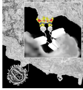
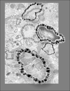
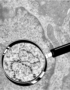
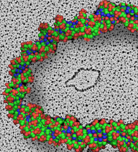
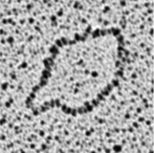
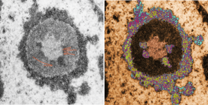
Shapes from biology (especially microscopy) sometimes are reminiscent of the most mundane objects in our daily lives. This nucleolus has reminded me of a donut with sprinkles for several decades.  In searching the nucleolus for clues to the connection between the sizes of apparent granule-fiber, beads on a string, things to what has been reported in the molecular structures online, I used this particular nucleolus (from a baboon that had been given perfluorophenanthrene emulsion (probably IV)) and some tissues biopsied. This is lung. In other views, this particular cell (not clearly identifiable but likely an alveolar macrophage) contains PFC droplets.

The “sprinkles” for this donut represent the dense fibrillar component (entities measured on a post from yesterday; the cake part of the donut is represented by the circular structure in the center of the side-by-side identical micrographs, which is the granular component. NB, the granular component on the left is light grey and round with the fibrillar center being the center of the donut.  It was noted in some of these nucleolar structures that there is such definite banding and that the lumps on a string is a very good casual description of the organization of RNP along a template and the measurements are pretty good.  As a reference point the diameter of a ribosome is about27 nm and the space between ribosomes in this particular cell is around 58 (or 2x the diameter of a ribosome). The diameter of the beads in the granular portion is 21nm, with a distance separating them of from adjacent structures is quite large and prominent, around 42nm (twice the diameter of the granule and though smaller in overall size than the ribosome and its space) is quite similar to it. The granules on a string in the dense fibrillar portion of the nucleolus is close to 30nm, slightly bigger than the average ribosome, and the space around these larger granules is the smallest among the three, at 38nm.

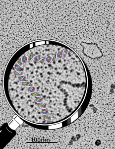
A second organization to the granular portion is seen in the parallel alignment and even spaceing of the granue. Orange lines lie over the very obvious linear pattern in the granular center of the nucleolus. A similar linear alignment is found in the dense fibrillar portion which becomes very very obvious in cajal bodies (not seen in this micrograph).

electron micrograph of the nucleolus of a baboon lung cell
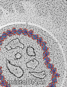
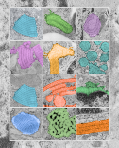
This image would be a fun cover submission…  maybe someday i will make up a panel with three or four TEM look alikes. I know i have a valentine, and a ghost and a donut.

Awesome aids virus and some factors

I made this diagram for someone a decade ago, whose name (LS) means nothing to me and the names of the protein complexes escape me. It was a cover submission for an article submitted to PNAS at some point and didn’t make the cut, and I am sure LS never paid me.  It has some interesting information in it anyway so I am posting it as an illustration.  This version is considerably modified so I am not breaking any copyrights…besides it was my illustration in the first place (LOL).

My kind of science art: Starry Night by Alex Parker

Wonderful images exist in science, I can attest to this having taken thousands and thousands of electron micrographs of the intricacies of the myriad cells in our living bodies. It is an amazing universe, whether tiny or distant. This particular montage by Alex Parker reminds me of my days as a graduate student in anatomy, where i cut up hundreds of black and white micrographs (those i printed (yes wet processing of black and white prints) into thousands of pieces) and pasted the them each, one by one, into a 4 x 3 abstract sunburst montage.  I don’t even know where that picture is at this point, being almost 45 years in the past.  But here is a fresh version by another graduate student.. Alex Parker…  how fun this new iteration of an old obsession — sorting, ordering, recreating. It would be nice too if this were his own abstraction of the cosmos or something he designed, but it is also nice to recreate the past with the brink of inquiry.

Alex Parker has some nice videos as well, here is one. and here is another (totally amazing asteroid animation)


More microDNAs for cover submission

I must have really made a lot of variations for this cover submission, here is another that I modified this morning.  It is a little confusing, but the cell behind is a has a large nucleus (left middle of the micrograph), which is in distress, with some indications of impending apoptosis, mainly the finely granular chromatin at the inner nuclear membrane, and the prominence of interchromatin granule clusters, and a rather large nucleolus (with prominent fibrillar centers and dense fibrillar components (in preparation for the trip down the apoptotic pathway). And on that image I superimposed a dozen or more of the images of microDNAs that Dr. J.D. Griffith gave me to organize into a cover. So the microDNAs, (of which three are enlarged bursting out in dimensional relief) that are mostly black and grey high contrast, while there are many others lesser in size that are superimposed in relief over the original transmission electron micrograph of the cell.

Electron micrograph of nucleus and overlay with microDNAs

Still working on a cover submission with Dr. J.D. Griffiths microDNA images, superimposed over one of my own electron micrographs of a cell with prominent nucleus and highlighted within that as an inset is an enlargement, over which is superimposed his micrographs of microDNA.

I don’t think this image was selected for submission, but it is pretty nice none-the-less. I am prompted to post these, after looking back over the archives (for other reasons).

DNA and microDNAs

See these images which were submitted, but not accepted for cover illustrations.  Electron microscopy by Dr. J. D. Griffith, and rendition by Dr. Marian L. Miller.  We were working on a cover submission, and those examples below are just a few that were organized, plus the one I liked best shown in a previous post on this  blog here .  The bottom right image I find this morning while googling, skimmed, and referencing some keyword advertizing company…. this makes me furious. The pasted up DNA is middle right, and all these are Dr. Jack D. Griffiths micrographs of microDNAs.  Shame on you skimmers and spammers and hackers.

..yes, one more cover submission illustration — I just love these granules

..yes, one more cover submission illustration — I just love these granules, and I think this might be my favorite yet. Colored all the granules in (using photoshop) overlying their own original images from the basic research on this putative SP-A granule, and arranged them in a highly ordered format (haha) which is certainly not really my normal MO, since I am kind of a random thinker.  But this is nice…in my opinion, and also a little artsie-craftsie for a scientist.. but thats what covers for scientific journals are for.