Category Archives: Nucleoar organization

nucleolar-organization, RNA translation transcription electron micrographs

Nucleolus and SV40 infection

Looking at the sum total of nuclear architecture seen from the viewport of the electron microscope, I am trying to summarize the topology and/or segregation of protein-DNA, protein-RNA and protein-protein domains therein.  It seems that many names have been associated with very many small structural entities within the nucleus, (and nucleolus), and not all these molecular studies have been diligent about identifying them with transmission electron microscope (actually using fluorescent probes and light microscopy gives pretty pictures, merging reds and greens — but this is still like looking at the barn door when one wants to see the insects on the wood) thus making it a little difficult to interpret their results. I did find an early manuscript, from 1963, which helps, published just about 10 years after the commercial availability of the electron microscope (yes I laughed since the author of the manuscript named the equipment, an old Seimens 1 — the microscope I used for 30 years was the next generation, the Seimens 1A.  yep, old).

nucleolus electron microscopy SV40
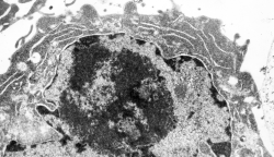
The publication is by Nicole Granboulan and appears to be readily available for perusal and it focuses on the nucleolus, a portion of the nucleus dedicated to ribonucleoprotein biogenesis. I have taken portions of three micrographs, an uninfected, and two SV40 infected, and put them side by side to show the changes in the nucleolus as it becomes a production site for viral pre-ribosomes as shown by the great increase in the granular area as time after SV40 infection increased. There is an augmentation to the dense fibrillar compartment, which initially to me looks like a crescent moon with the fibrillar centers poking in like mushroom stalks, to feed the production of viral RNA.  It is clear that the surface areas between the fibrilar centers (where the rDNA genes are located) and the inside of the crescent moon portion of the dense fibrillar areas has occurred as well. An increase in the granular component in uninfected nuclei (uninfected cells) is also occurring when cells are in S phase of the cell cycle.

The black and white diagram overlays with white arrows show the change from U to M shape of the dense fibrillar regions, and the white areas of the cartoon, being the fibrillar centers.
Fibril, filament, snRNPs, granular component etc

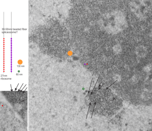
This micrograph I am using to determine the sizes of the different nuclear structures, so this is the first attempt to define at least four different sizes of components. For me, there is similarity in the size of the granules (beads on a string maybe something like 30 nm) in the cajal body and in the nucleolus. Arrows point to the kind of layered banding parallel strand look of the cajal bodies,  the red circle is about the size of a cytoplasmic ribosome (which would be something like 27nm) and the diameter of the rounded object beside the cajal body (something on the order of 60nm) and an even larger fibrillar component seen through out the fibrillar centers and also just a little above center left (orange spot) which might be 130 – 150nm.  So there are four measurements of fibrillar components in this nucleus, so far.  Will post more.

electron micrograph liver 14CoS ko nucleolar architecture

Mono ribosomes

Cytoplasm of an HeLa cell grown in vitro, no UV exposure, but inhibitor of caspase 1 added.  Lots of monoribosomes in the cytoplasm.  So this is still part of a study to summarize the ultrastructure o the nucleus, nucleolus, in apoptosis. This nucleus has a very large nucleolus, large fibrillar centers, not that much dense fibrillar component but a large granular component. Not a great micrograph, but data, none-the-less.


Interchromatin granule clusters: HeLa cells UV + inhibitor of caspase-1

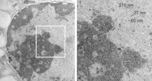
Interchromatin granules (that is a cluster of granules) of varying size, but mostly rounded areas, are found in the nuclei of mammalian cells. These are clustered in the nucleus, especially in and around apoptosis as seen in the lower magnification electron micrograph on the left below. They are, according to concensus, composed partly of pre-mRNA splicing factors.  They are not static structures, and are active sites of transcription, size can be several microns or hundreds of microns in diameter.  They were found in a study done years ago, to be really prominent in late phases of apoptosis. The image on the right shows particularly well (though the micrograph is not sterling by any means) that they are composed of 20–25 nm granules that are connected like beads on a chain by thin fibrils (also visible).  The red dots in each of these two images (the right image enlarged from red box on the left) are the size of ribosomes, and the bar markers then are 10 times bigger (270 nm).  There are at least 3 other interchromatin granule clusters in the micrograph on the left. That shows the beads in the right hand enlargement to be just around 20-25 nm in diameter (something a little smaller than the accepted middle size of a ribosome (27 nm) (selected from the same micrograph.

Interchromatin granules may be collections of snRNPs and other RNA processing components involved in producing mRNA. These granules are so evenly spaced as to be a marvel.  neg 14138_block 61002 HeLa cells, UV and inhibitor of caspase 1. (BTW, dont ask what the dense areas to the right of the interchromatin granule cluster is…. ha ha)

rRNA transcription just confusing

This is so awesome, just read part of this article by cheutin et al_awesome nucleolar diagram and view the TEMs and diagrams, were that I were beginning a career in TEM tomography, i would be in heaven. But 40 years too early.  So at best, is to take the new information and correlate it with studies from the past, making use of the data that I have and thousands of electron micrographs to play with.

TO begin with, 14CoS nuclei and nucleoli with some bar markers, just getting acquainted with the possibilities for labeling these structures. So here is the image from a previous post, and the segment enlarged with the two cajal bodies, tiny red circle is the size of a ribosome. Nucleolar structures I have not measured yet, but according to the publication by Cheutin et al, linked above, these will be similar and important in the pre-apoptotic hepatocyte nuclei found in this mouse line when they are not rescued by administration of oral 2-(2-nitro-4-trifluoromethyl-benzyol)-1,3-cyclohexanedione (NTBC).

electron micrograph liver 14CoS ko


Nuclear pore: canine type II alveolar cell

nuc-pore__dog_type_II_cell_nucleusThis cropped portion of an electron micrograph of a nuclear pore was taken from the nucleus of a canine type Ii alveolar cell. It shows the filaments on the cytoplasmic side, and a bunch of ribosomes positioned just outside the nuclear pore, and two very distinct structures, about ribosome size that are either coming or going through the nuclear pore.  It made me laugh, as it almost seemed as if they were going or coming in a “pin ball” fashion, being shot through the pore directly in the center. Do you see both of them? one is almost dead center of the micrograph, abd just elow the nuclear pore, and one is almost directly below that.

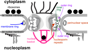
This electron microscopic image prompted me to try to figure out what is known about the nuclear pore complex in general, and every diagram I found in the literature was just a little different, even that found on wikipedia had some elements that I could not assemble over top the micrograph (which is basically what i wanted to do), so the diagram which accompanies this fine structural view (ultrastructure) is way off… I would love to find the time to put together a better diagram.

nuc-pore_diag