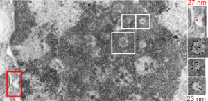
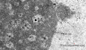
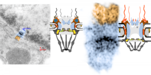
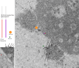
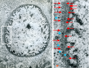
I was looking at this micrograph of a n hepatocyte with the fibrillar centers and dense thick fibrils in their midst, and the granular zone around the outside of this nucleolus. The latter being very punctate in appearance, just really obvious. I measured these once again at about 23 nm in diameter and noticed that around many of the fibrilar centers where was a basket, or ring, or radial distribution of these small punctate appearances. OK so they are not really punctate, but can appear smeared like fibrils too, though the granular appearance and the nomenclature is well entrenched in the literature, they are just going to be strands not dots. It could be that these radial dots around the fibrillar centers are bands, or windings at a given distance apart, kind of like fingers of a basket, or more aptly put, like a cage, with wires of 23 nm in diameter.
Electron micrograph here shows a portion of a nucleolus with three fibrillar centers cut out and enlarged (original sites are boxed in white), and a portion of the outer nuclear membrane with ribosomes (for measurement – one ribosome being about 27 nm in diameter, top right image with red dot and text). The cross section of the cage, or basket filaments around the fibrillar centers are shown as black dots.
