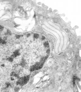
Going through additional species of animals looking for possible corollaries for the intracisternal bodies found in the RER of guinea pig, ferret and dogs, I examined just a few transmission electron micrographs of rabbit. One animal from a study with Leland Clark Jr, very likely involving artificial blood, and four other rabbits in studies with David Warshawsky, with carcinogens (likely dibenzcarbazol, and also iron oxide). In the former (see the unretouched, but contrast enhanced micrograph below) I did find three stacked RER cisternae which had the look of some of the cisternal bodies found in three species but I was not able to detect any of the layering–aka—periodicity within these cisternae. Well actually i was tempted to see it, it seemed to me that very faintly there was some tendency to see banding. You can judge for yourself. Top micrograph.
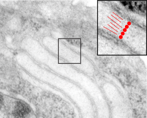
In the bottom micrograph at the thinnest part of the cisterna (inset marked by a black rectangle and enlarged to upper right corner) at a near perpendicular cut, it seemed to me that the cisterna was more than 100 nm in width. (A ribosome adjacent to the cisterna was used as a measuring stick, the latter presumed to be 25 – 30 nm in diameter; red dots). What might be construed as linear patterning, or parallel periodicity, appears as red lines.


