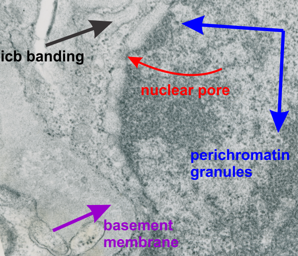
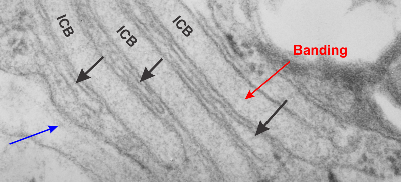
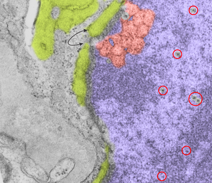
Looking at an electron micrograph of a guinea pig alveolar type Ii cell, (my guinea pig number is 301 – block 17082) archival material from the 1980s, I found this really interesting pattern in the nucleoplasm just above a nuclear pore. On either side of the outer nuclear membrane spanning the nuclear pore was the granule protein stuff that I have been working on, trying to place it possibly in the surfactant protein family, but being overproduced to the extent that it becomes its 18-mer, in mirror and/or vertically flipped position, times 2… producting the 100 nm banding that I see. But for this nucleus, just to the right of the nuclear pore marked with a two headed arrow pointing to the lime green intracisternal protein within the RER, there is a pattern that just “popped out” to me which is quite large (relative to the size of a ribosome…. and NOT to be confused with the hexagons in a tangential view of a granule but in the NUCLEUS proper) and sort of hexagonal in shape (which is what one expects when pressing together spherical bodies which I pseudocolored a salmon peach. Red circles surround structures that look like perichromatin granules (I am sure I could have found more).
Daily Archives: August 19, 2016
Intracisternal granule from alveolar type II cell of the guinea pig
Nucleus, other organelles, nuclear pore, intracisternal protein accumulation, perichromatin granules, and part of the basement membrane are labeled in this electron micrograph of a type II cell from the lung of a guinea pig. Nucleus is on the right and a very thin band of cytoplasm is present, then there is the extracellular space (with basement membranes and parts of other cell processes). Basement membrane – purple arrow and label; red arrow and label – nuclear pore; black arrow and label – intracisternal protein which might be surfactant protein A (overproduced). Perichromatin granules (adjacent to heterochromatin) are shown by blue arrows and label.
Smooth ER between RER lamellae in guinea pig alveolar type II cells
This electron micrograph shows three very long (a portion pictured here) lamellae of RER with a surfactant ?? protein, maybe surfactant protein A accumulation which shows a very very light area of banding – ICB) , and sandwiched inbetween these three are flattened cisternae (black arrows) which do not apparently contain the protein accumulations that is present above and below each. Whether these flattened cisternae without apparent protein are going to be sectioned down the block as thickened lamellae with protein content, is up for grabs, but likely (see lowest black arrow which points to a thickened part of the flat cisternae). This configuration is a little unusual. Other things to observe in this electron micrograph are: part of a lamellar body – upper right; tiny hint of the central band typical of what I think is overproduction of surfactant protein A – red arrow; basement membrane beneath the alveolar type II cell, and what is probably an endothelial cell – blue arrow;