It is a common problem for scientists to be unable to communicate ideas, problems, discoveries, hypotheses and conclusions to colleagues and laity. I think it is because there are so many levels of “knowing” something. Not all people can translate knowledge into pictures. That sounds like a silly statement, but personally, there are many ways that I “know” the cells and tissues that I work with – but mainly I know them “visually”, and I have many many colleagues who “know” cells in a different way, and really have no clue as to whats going on structurally, or where.
I see this disconnect when one of my colleagues says something like – “we found this in liver” or we see this in “skin” or the changes are in “lung”…. what!!! that is completely wasted talk, there is no information in those statements, at least for me. I think of liver as a mix of parenchymal cells, supporting cells and systems (venous and arterial and neurological), matrices and architectural components, secretory and excretory elements, immune and detoxication apparati, protein production, carbohydrate storage, not to speak of all the myriad and wonderful ultrastructural components of each cell type, such as hepatocytes, duct cells, endothelium, that most individuals have never seen. I could go on and on…. in all humility…. I know nothing about liver, but apparently, neither do they. There is no useful information in the word “liver”, at least to me. Saying “liver” is like saying “someday” to a person who is asking for a specific point in time to be declared.
Similarly, after many years of working on the mouse back skin models of carcinogenesis I began to dread hearing others talk about “skin”…. Skin is a complex organ system, and conservatively speaking, has a dozen unique cell types, and about as many functions known to date, and many many more unknown. This has come to be true of every organ and tissue and cell type I have looked at through the electron microscope. And just for the record, in so many ways, cell cultures just don’t function like, or look like, cells in situ, that I just cringe when the parallels are drawn. Yes, I confess, we all learn from reducing systems to their lowest common function… I understand that.
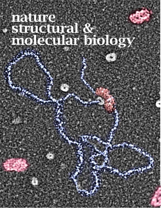
So the bottom line here is that a “careless use of words can cause confusion BUT a careless diagram causes 1000 times more confusion“. Shall I list the ways the image below causes me grief.
- The alveolar type II cell is not a box, though diagrams giving it a cuboidal shape are usually acceptable, this one is not. The alveolar type II cell (as this box doesn’t represent (below)) has distinct plasmalemmal portions which have very different functions and morphologies, e.g. the apical (microvillar) area that interfaces with the alveolar space, and the basal or plasmalemmal side adjacent to the basement membrane, and the cell-cell lateral membranes as well, none of which is pictured or identified in the diagram below, thus creating a false impression of the cell membranes. In fact the lamellar body pictured inthe diagram is positioned such that it looks closer to what I would be inclined to assume is the lateral plasmalemma, than the apical membrane, which is completely erroneous since lamellar bodies are exocytosed from — and surfactant components are recycled from — the apical membrane.
- The nucleus here is completely wrong. There are no breaks in the inner and outer nuclear membrane, nope, and the only punctuations in the nuclear envelope (composed of the inner and outer membranes and the perinuclear space) comprise nuclear pores, which makes the nuclear membrane continuous not dashed. No breaks as pictured below. Yes the perinuclear space and outer nuclear membrane are intermittently continuous with the RER, this is true, but I have never in 40 years seen a stretch of continuous RER that almost completely encircles the nucleus as alluded to in the diagram just below the nucleus… this is careless, and false information about alveolar type II cells where the RER is normally short flattened cisternae.
- It would be wonderful if when diagrams like this are prepared for publication if someone made a decision to either use 3D or 2D and stick with it. (Similar issues are seen when in writing a manuscript, the author jumps from present, to future, to past tense..ha ha) Case in point: the cylindrical objects for two transport proteins are picture in something that is supposed to be 3D (no attempt at using a structure that might actually look something like the protein they are depicting) and 2D for the remaining structures. They give prime time to the one transporter for phosphate and this erroneously gives the impression that this is the only channel, transporter, or pump that is relevant in type II cells homeostasis which is totally wrong. And even if one declares it was done for simplicity’s sake, this doesn’t hold water since the disease that a malfunction of this sodium/phosphate transporter protein might produce is quite rare (according to the authors) so to give it so much press seems silly.
- It has been my observation that the nuclei in the type II alveolar cells is more central and basal than off in the corner, so this is misinformation as well.
- The appearance of lamellar bodies can be close to the base of the cell, on the sides and also in the apex, but clearly the exit is from the apical membrane in all likelihood so the position of this lamellar body might have been better placed nearer whatever membrane portion was supposed to represent the apex. Judging by the blue arrow, pointing to the electron micrograph (which could have been much much larger for clarity) shows the lamellar body exiting the cell in the lateral membrane, which of course is wrong.
- When diagrams of lamellar bodies are made, one could quite easily approximate the “real look” of lamellar bodies, which as the name suggests contains many many lamellae….and often is as much “empty” space as lamellar space, so why for goodness sake is this lamellar body shown as a solid object? That is just missed opportunity to provide useful and accurate visual information, rather than the information that is portrayed…. which is just inaccurate.
- As for putting in the diagram the golgi apparatus, (yep i should have used a capital for the G in golgi, as identified in 1897 by Camillo Golgi, but too long ago to be a relevant decoration) as the ONLY pathway for proteins from the RER and the only pathway from golgi to multivesicular bodies is much too restrictive. Mature lamellar bodies can be found in all quadrants of the cell, though some propensity exists for larger more mature lamellar bodies to be found at the apical membrane, many are found laterally and basally, which then renders the diagram below, inaccurate once more.
- A small comment, Type II alveolar cell, vs alveolar type II cell…. a little backwards in terms of convention. and I have done it as well, but stating the organ or tissue location is a good idea before naming the cell subtypes, especially since there are other types of type II cells. In a quick google search 53 occurrences of alveolar type II cell, vs 3 (one of which is mine, sadly) of type II alveolar cell, and quite a few times, type II pneumocyte, and sometimes lung type II cell. Convention is not always good, but in this case probably would have been nice.
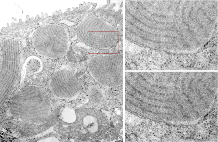
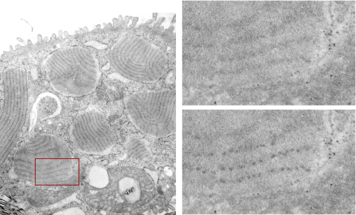
- The long intracellular distance the lamellar body has to traverse to exit what should have been the apical membrane (but here the exocytosis will occur on the wrong side (laterally) in this diagram and likely an inadvertent choice for the position of the arrow) is not consistent with what is seen microscopically, in addition, the electron micrograph in this diagram (the tiny black and white picture to the right of the box-cell) is tubular myelin, which in this particular instance is a very unusually large amount that is not often seen that way. This is a false indication of what surfactant actually looks like in the alveolar space of the cell fixed for fine structure…. in fact in these researcher’s own publications, surfactant with NO surfactant protein A, has NO tubular myelin at all, yet they reported no respiratory distress…. thus the image is generates false information while making a point. The scrambled looking surfactant more typically seen, with only tiny amounts of tubular myelin, is still perfectly functional surfactant, and the unorganized stuff is actually much more abundant in micrographs than the would be assumed from the tubular myelin inset below. I am not sure why one would even want to suggest that all surfactant looks like tubular myelin.
- Just to the right of the tubular myelin is a cartoon of a lipid bilayer. That diagram is most typically used to represent a cell membrane (plasmalemma) and in this case it is repeated…a large and a reduced version…. what? First of all, this lipid bilayer is not in any way what the lamellar and tubular myelin surfactant looks like. That lipid bilayer doesn’t have any proteins attached…. functional surfactant (by these authors many publications) has protein components, partly for structure partly for function, partly for other purposes (breakdown, recycling, innate immunity, others about which I do not know — but they probably do). The question is “why use this model of a cell membrane bilayer as a model of “what” do they really mean this should represent surfactant within the alveolus? There is so much misinformation in that depiction I can hardly tell what the purpose of the diagram (and its miniature friend) was.
- OK, here is an odd comment, but what was the purpose of the red arrow from the alveolar space back to the multivesicular body? And what was the purpose of the word “catabolism” — just silly redundancy. Who in life doesn’t understand the word recycling? Doesn’t that suggest taking apart and repurposing, AND to suggest that all surfactant is recycled through the multivesicular body…. yikes, there is a real leap, that cannot be substantiated by any electron micrographs of alveolar type II cells I have ever seen. I will have to check the position of multivesicular bodies (I won’t take time to quantify any results or create a dataset, but just from my observations, MVB are neither sufficiently frequent, nor sufficiently large to accommodate the volume of turnover, though they repurposing of some components of the uptake of surfactant from the alveolar space might well get shuttled off to MVB in those low amounts.
- So if the Abca3 transporter moves DPPC into lamellar bodies, this diagram suggests that it does so from the cytoplasm (but I think that this is moved through the membranes. OK, more later.
- OK, so i am bored of this now, I am certainly grateful that they didn’t deem it necessary to add the diagram of an alveolar macrophage. That might have triggered another rant. Best to all these authors, each of whom I have met. But this was a great exercise for me, and a wakeup call to all you scientists who think that you can carelessly slap together a box and a few ovals and a label or two and do any one any favor. This was clearly only accepted for publication by reputation, not by information.