Daily Archives: September 13, 2017

Awesome IPFD crystals in lysosomes

Perfluorodecyl iodide was examined once upon a long time ago as a possible PFC for blood substitutes. There are a couple of tissue blocks which I am re-examining to see whether there is a periodicity to the lysosomes that are included in these amazing phago-lysosomal structures, left over footprints as it were, to the presence of IPFD in macrophages (Kupffer cells, fat storing cells?, circulating macrophages, in the liver).
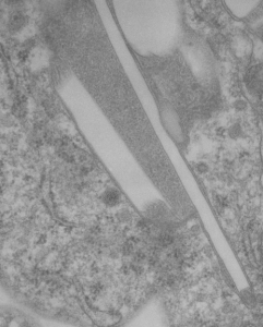
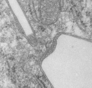
perfluorodecyl iodide transmission electron microscopyperfluorodecyl iodide transmission electron microscopy
This lower micrograph has very definite structure at the ends (one top one bottom) of the crystals. It is a little unfortunate that at the time i printed these negatives (back in the ‘wet darkroom days’ that I didnt use a finer grade paper and developer. Too much grain, but the little indents and rounded areas with a punctate density are going to be relly interesting. I think the lucent areas beside each will measure out the same as some fine lines that I see at the ends of other crystals.
Comparing perfluorocarbon emulsions: Clark et al, 1974

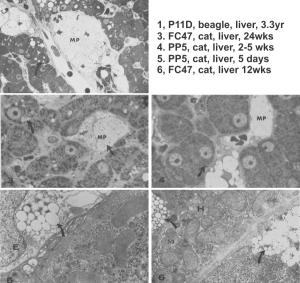
I ran into this publication that I had not even put in my own folder of pdfs, that was published back in 1974, Clark LC, JR, et al, in Microvascular Research. I was sort of surprised to see how much effort went into the assessment of perfluorocarbon blood substitute emulsion particles in the livers of several cats receiving either 1) no emulsion, 2) PP5 emulsion or, 3) FC47.  Early on it was evident to me that the re-emulsification of fluorocarbons in vivo was quite dependent upon the chemical characteristics (but i dont know which) of the PFC, combined with — or prompting the lysosomal response, coupled with the apparent diffusion and aging characteristics of particles within phagolysosomes.

I have extracted the micrographs from this publication and added the descriptions (which were unfortunately difficult to find) from the publication which shows that P11D and FC47 clearly differ from PP5 in the responses they elicit from the phagocytes in the liver (likely Kupffer cells and fat-storing cells, are RE cells. Top three images are light micrographs, bottom two images are transmission electron micrographs.  No bar markers unfortunately but in the top three use a nuclear profile as about 15 microns, in the bottom two, use the short diameter of a mitochondrion as about 0.2 microns.

perfluorocarbon emulsions liver transmission electron microscopy PP5 P11D FC47In this publication Clark notes that PFC ring compounds with O, and N, and straight chain PFC with O, hang around for a more extended time than those with just C and F.