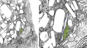
This is an interesting perfluorochemical (IPFD) and in the mid 1970s it was used as an emulsion for studies on artificial blood (blood substitutes). It was the only perfluorochemical used that had a crystalline structure in vivo, and while i have posted previously on it, i have hunted up about 10 negatives (i think the only negatives i have) from those long forgotten files and will examine whether or not there is any peculiarity about the enzymes, perhaps even a periodicity to the lysosomal enzymes which surround the IPFD inclusions. I have to assume that the cell in which the crystals are appearing is either a kupffer cell or an hepatocyte, I can’t be sure which.
Just from the beginning, here is an image scanned at low ppi, but it shows a curly look to the lysosomal structure surrounding this narrow IPFD inclusion. IPFD is purple outline, green is the lysosomal contents. Image on right is enlarged from left.
