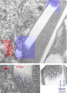
I looks like the lysosomal enzymes at the end of the IPFD crystal inclusion-structures might be tubular. But then as the enzymes pass away from the IPFD itself, there can be a coiling up of the protein with less rigidity. Measurements at ends of IPFD look smaller than those in the lysosom proper… may be error of just having only measured a single crystal. Same negative and block as previous post, and the ends of the IPFD crystal are colored bluish, and the area measure for ribosome size is in red. Enlargements below, with areas specific measured highlighted. The dense part of the period and the lucent areas beside it.
Daily Archives: September 15, 2017
Texture comparisons: background, cytoplasm and IPFD-lysosome
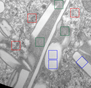
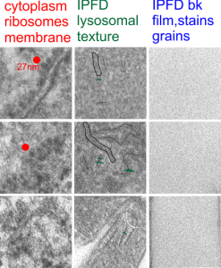
I am trying to determine whether there is a predictable texture to the proteins within phagolysosomes which contain IPFD (perfluorodecyl iodide)(which I am positive there is–so this is academic). I have compared territories from one micrograph with marked inset boxes: red boxes=cytoplasm with a ribosome size marker for comparisons of texture-size); green boxes=portions of the compact and textured lysosomal proteins found just within the limiting membrane of the phagolysosomes which contain IPFD; blue boxes=the background texture of the micrograph found over the “empty” IPFD granules which consists of the acetate film grain and the stains– osmium, uranyl acetate and lead citrate grains. No photographic paper grains are present as this was scanned at 5000 ppi from the negative.
I measured a similar texture pattern from the end-on portion of the lysosomal proteins in last post, and dimensions are similar but not identical… likely my fault, not the fault of the proteins. So todays measurements are something around the following: dense areas at around 17nm (corresponding to the “spot” in yesterdays post which was around 13nm) and the surrounding more electron lucent space measures about 34nm (which in yesterdays post was closer to 40nm)–all likely resulting from variations in how i measured ribosome size, which is the benchmark.
Image below is from a randomly selected phagocyte in the liver (I cannot identify whether this is a Kupffer cell or other phagocytic cell type). These cells can be closely aligned with sinusoids but also near portal tetrads. They are typically “overflowing” with IPFD particles. IPFD stays around for months, unlike other some perfluorochemicals.
Neg 9722 block 3775 10% IPFD 5%F68 injected 8 14 1973 100cc/kg sac 5 8 1974 mouse liver 267 days.


