I worked hard and lobbied continuously for this cover. Here is a link to the journal in general.
Daily Archives: September 18, 2017
Substructure of the lysosomes containing perfluorodecyl iodide
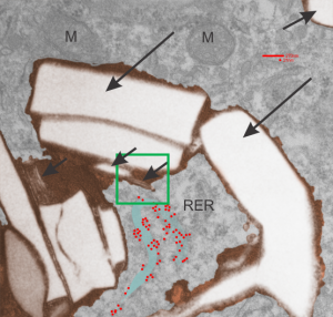
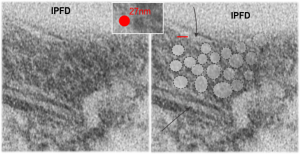
Substructure of the lysosomes containing perfluorodecyl iodide is very interesting. There are issues both with the lysosomal membrane, which in some places may be continuous with other membranes, in particular, i think maybe the RER. Because perfluorocemicals are kind of “slippery” and I think they can travel easily retrograde-style back up into the RER, then it makes sense that some profiles of RER would be seen at sites within the lysosome periphery and the adjacent cytoplasm. I haven’t been able to equivocally find ribosomes on the actual lysosomal membrane to verify that concept. There are perfluorodecyl iodide tiny crystals…. this is interesting…. an obvious type of fracture or separation plane along the long axis of the larger crystal inclusion certainly is evident…. and i am more convinced that the pattern of lysosomes (a dotted or tubular pattern) is on the order of 20-30nm diameter or width.
Here is an electron micrograph: mouse, neg 9722_10%IPFD, 5%F68 infused 100cc/kg, 8-14-73 sac 5-8-74, 267 days post infusion; liver. Brown areas=lysosomal enzymes within a bounding membrane, white=IPFD, red dots are ribosomes; pale green is cisterna of RER, green box is area enlarged below.
So the bottom two images, left unretouched, right unretouched but with translucent overlays where I see a pattern within the lysosomal enzyme membrane. Arrows here point to fractured off crystals of IPFD, and the red line is a marker for 27nm which is the same size as the ribosome measured (top inset)…. this makes me wonder if there is some connection between this pattern and lysosomal contents.

