Monthly Archives: October 2017

This is why we hire translators

While looking up some info on another perfluorinated compound used in testing for that group of chemicals’ usefulness in the field of blood substitutes I found this cute description — which just made me laugh a little.  While admittedly, I cannot speak another language (except just enough french to get to the restroom), I would never make a company product listing without having someone sit beside me and personally help me translate.  Otherwise something like what is listed below would happen.  Hey, maybe google did this translation, haha.

“Fluorinated fabric finishing agents and fluorinated surfactant intermediates. Development and production company perfluoroalkyl iodide, perfluoroalkyl ethyl iodide, perfluoroalkyl ethanol, perfluoroalkyl vinyl and perfluoroalkyl ethyl acrylate and other products is an important chemical raw material, can be transformed to the corresponding alcohol, thiol, and sulfonyl chloride such as various fluorine-containing intermediates for further synthesis of various fluorine-containing surfactants, fluorine-containing finishing agents and other fluorine-containing fine chemicals. Such fluorinated products with high thermal stability, high chemical stability and excellent performance hydrophobic hate oil, with its made of fluorinated finishing agent with conventional textile finishing agent incomparable characteristics, through their treatment of textiles with a variety of excellent performance, and thus much attention and welcome from domestic and foreign markets, to become the mainstream of today’s textile finishing.”
Phagolysosomal and crystal patterns in IPFD inclusions in liver

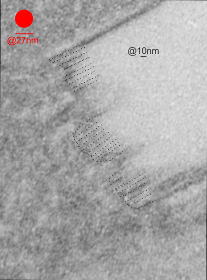
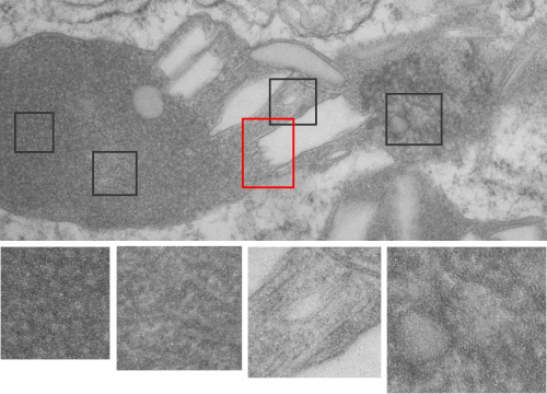
Two prominent areas of patterning are apparent in phagolysosomes in liver after IPFD infusion. The IPFD crystal itself, and at least four distinct protein patterns in the phagolysosomal proteins. The first image below is an enlargement of the red rectangle, and I have marked off with lines some of the pattern at the long end of the perfluorodecyl iodide crystals which seemed to work out to about 10nm in the short dimension. 23 measurements made at the end are marked off with dotted lines. A red dot=the dimension of a ribosome taken at the same magnification for reference.  Below that is the low magnification image (unretouched — I removed a scratch from the top image but no data were altered as it was not in the crystal itself). Black boxes correspond to four very distinct protein organizational patterns within a single phagolysosomal body which also included the crystal in the top image. I am pretty confident that these patterns are a clue as to which lysosomes are present, and 4 may not be the only ones which show oligomerization into ultrastructural patterns. One thing to think about is that the kupffer cells (likely) in liver acting as macrophages which engulf (and hold onto for months) IPFD crystals will have a different panel of enzymes for lysosomes (and peroxisomes) than hepatocytes, and seemingly can become multinucleated under the conditions of massive IPFD inclusions.

IPFD: phagolysosomes with protein patterned substructure

Phagolysosomes which include IPFD – 1 iodoperfluorodecane aka perfluorodecyl iodide have some interesting lysosomal enzymes.

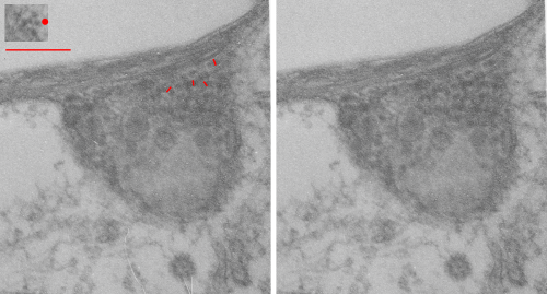
Of these many enzymes I am thinking that there are many which are “folded” or oligomerized, that is those phagolysosomes which have IPFD show a heterogeneous substructure, zones of tube like enzymes, and areas of mostly untextured protein and then areas of unique coiled ultrastructure.  Below are two electron micrographs, the one on the left unretouched scan of a print, and the one on the right retouched (though I can hardly tell the difference – the one on the right had the coiled nature of the protein burned just a tiny bit with photoshop and several pieces of lint minimized with the bandaid tool in photoshop but no data were changed).  Red dot is ribosome (taken at about 27nm) and the thickness of one of the fibrils is something under 25nm…  which is a little larger than i measured before at 20nm but i am probably responsible for the variation), red bar is 270nm, short red lines are measurements of the diameter of the coiled (polymer or oligomer protein), pale untextured area at the top of the micrograph is the footprint of the IPFD crystal, the whole area is mostly phagolysosome. Inset is the cluster of ribosomes used for size.


Peroxisome in mouse liver after perfluorodecyl iodide

Peroxisomes: I have learned a few basics about these organelles. They are an old organelle evolutionarily, and the enzymes within have become unique to the organisms, species, and cell types wherein they function.  So in the case of the liver of the mouse, there are going to be many enzymes, but basically for the following purposes: 1) for directing peroxiome-relevant proteins produced by the RER into two places, 1) including the peroxisomal limiting membrane, and 2) the peroxisomal matrix.  Matrix enzymes would include those that are protective, in that they metabolize reactive oxygen molecules, and participate in fatty acid metabolism, and peroxisomal matrix those enzymes that have evolved with the species and its environmental pressures and xenobiotics (which would