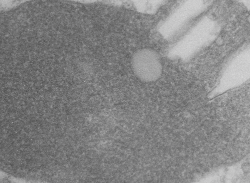
I have cropped three boxes from the image and changed the contrast (75Lysosomal response to the presence of perfluorodecyl iodide is kind of unique. After a 3 month hiatus (working on putative SP-A granule) it is nice to be looking at these crystal structures. I think I have posted this lysosome before, but am posting it again in reference to the three zones of texture and electron density that I am pretty sure I can see in this particular granule (not including the perfluorodecyl iodide crystals themselves). I took three areas of the crystal and changed the contrast (up 75 in photoshop) and also various levels of brightness to see whether a manipulated image would enhance any pattern present. So beginning, and certainly an obvious “overall density” change exists in these lysosomes. In top figure below, red dots represent noise . Below that, the differently contrasted images were vectorized using the same criteria for each so a difference in hos this renders beings more credibility to thought that there is a difference in protein density and likely protein organization in these separate crops from la single lysosome. The pattern, not yet found, just a hit that it exists.
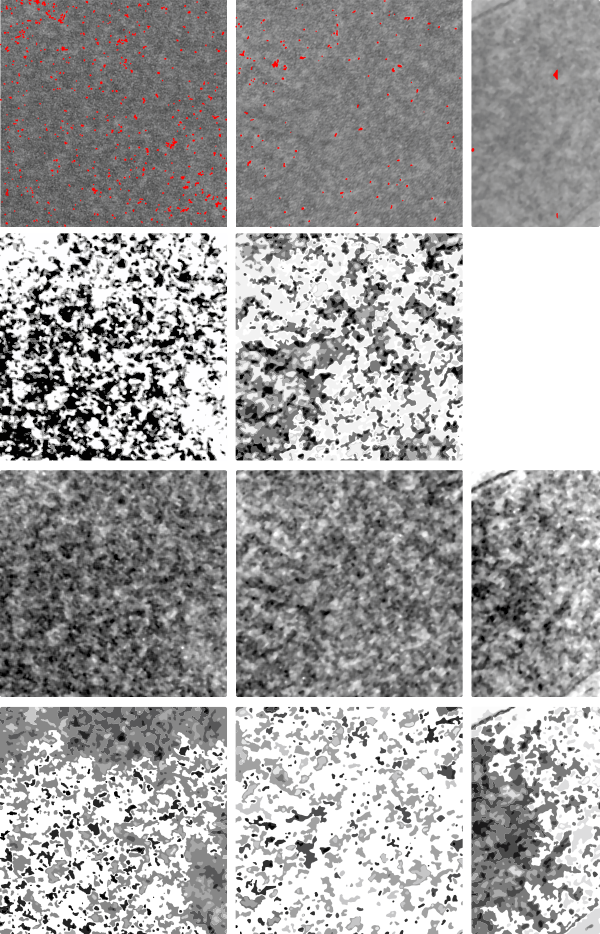 if you are diligent, you will be able to match the textures to the exact places I cropped (yep i should have made boxes on the original so you can easily see them), but i can see the pattern in the images below and where they came from on the original top micrograph without the need for boxes.