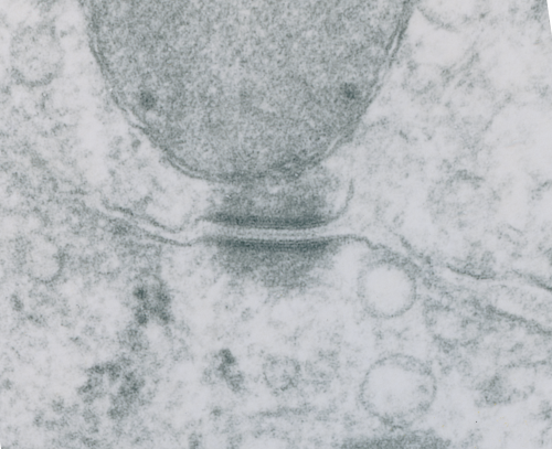
Looking still for some molecular models which fit the images I see for desmosomal mitochondrial junctions (adjacent structures tethered by intermediate filaments). These junctions are so prevalent that I cannot understand why they are not part of the everyday histology lessons. This particular mitochondrion and desmosome are from a mouse liver (control for other studies) . The sectioning and orientation are no too bad though it would be nice to have even more detail (haha as much detail as one might get from electron tomography, which isnt going to happen for me so i will do the best with what i have).
The molecular models of desmosomal proteins, which are pretty widely published, may have relevance to the structures here. The first think I am pretty convinced about is the stretchy hair-pin zigzag like connection of the cadherin molecules within the center of the desomosomal structure, and the little periodicity that is apparent (10-14nm spacing gets calculated from this image). I found a nm marker only on the molecular model for intermediate filaments which was 5nm (a very helpful marker indeed) and it comes close to confirming what I have as the magnification on the micrographs as judged by a 27nm ribosome standard. The proteins overlying the TEM here are just forced to size, as there were no nm markers on those models… and the position of the one large molecule (desmoplakin) is off to the side waiting to be figured out). There are big gaps between what I see with TEM and what the molecules say…. it is fun to try to figure it out.
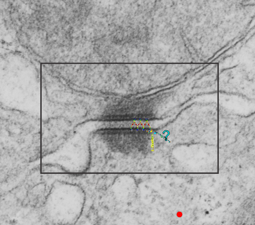
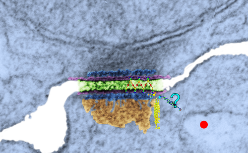
Upper micrograph, unretouched, also with rectangle for enlarged and pseudocolored image below. The bottom image has the intercellular space colored green, and you can see the cadherin springy wishbones above it, and also the plasmalemma and likely other proteins (plakophilin and plakoglobin) sort of carelessly put in place, and the desmocolin off to the side in an “unknown” placement. Red dots are ribosomes (for size reference).
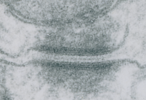 I looks in this image like the plasmalemma of each cell has a very rigid separation of the two leafelets.