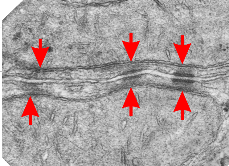
Both sides of two adjacent hepatocytes show two mitochondria each linking to three desmosomes. Nice to see such symmetry and the implications are quite amazing, in that whatever is happening at the plasmalemmae of these cells is happening “together”, really nice intracellular communication. Red arrows point to the inner mitochondrial membrane, just above where the intermediate filaments of the desmosomes approach the outer mitochondrial membrane. I wish i could say that I saw some cross sections of intermediate filaments here, but i didn’t. The double desmosomal mitochondrial junction on the left is more tangential than the one on the right. The pair in the middle is intermediate.
Daily Archives: December 6, 2017
Intermediate filaments tethering desmosomes and mitochondria
I have mentioned this phenomenon before but i keep finding images which speak to the prevalence of mitochondria (in this image two mitochondria are tethered to a single desmosome, one each in two adjacent cells). in addition to the fun double tether here, look at the “spots” which round the corner of the smaller mitochondrion…. tiny dots of what I measured as about 12nm, but the intermediate filament on cross section is 8-10nm, therefore my guess is that these wre cross sections of intermediate filaments. I did enhance these dots in any way except to change the contrast of the lower half of the image using photoshop. The dots ” cross section of evenly spaced intermediate filaments” are very prominent on their own. Pink dots are proximate to the filament x-sections, the red dot is an approximate ribosomal diameter. 30nm bar in middle is about what the desmosome measures, oriented perpendicular to the two plasmalemmae. N 6105 Block 5080 Mouse, female, orig mag 36000