Desmosomal symmetry: not a complete mirror images side to side, since it seems to occur in my micrographs, and obviously too in this micrograph from Green and Gaudry, that the separate cells (adjacent cells bound by the single desmosome) may have a propensity to have intermediate filaments coursing by at perpendicular angles. Maybe also chance – I guess it is a 50 50 chance to be chance (haha).
But also in this quote from Green and Gaudry.…I take exception to their remark that the desmosome is tripartite. A quote from their paper yielded this text of which several parts make no sense to me “electron micrograph further illustrates the highly organized ultrastructure of a desmosome (yes, i agree) in which mirror-image (partly agree, but not always true), tripartite electron-dense plaques (show me 3, ha ha, and are you counting lucent bands, plasmalemma (not really to be counted and intermediate filaments?) sandwich a central core consisting of adjacent plasma membranes (wait, plasmalemmas are NOT part of the central CORE, and what is meant by CORE anyway, that is a term that needs definition) bisected by an intercellular zipper-like midline (yes, zipper like dense midline -i agree)
They didn’t take into acount the different directions (which may be purposeful) of the intermediate filaments coursing by the desmoplakin molecules in the  adjacent cells… as sometimes they appear as 10 nm cross sections and other times as low arcing swooshes. I ask, what part of the plaques are divided into 3??? This is a confusing, since there is nothing distinct about the 3 parts they refer to… they could represent many/or any different layers of this organized structure. Their own electron micrograph (pasted below) shows pretty convincingly that there is not total mirror symmetry to the desmosome since their cell on the bottom part of the image shows cross sections of intermediate filaments while the cell on the top part of their image shows the longitudinal swoosh of intermediate filaments.
One thing that their electron micrograph shows that is rarely commented on is the desmosome annulus… this ring which of plasmalemma which is just slightly morphologically different than plasmalemma further from the desmosome.
While on the topic of their diagram, i think more care could have been extended to the depiction of the tight junction which is really a weld, and they show it as a wider structure than the two blended plasmalemmal membranes really are. And the adherens junctions are diagrammed to be as prominent as desmosomes, which they are not, and the labeling of adherens junctions actually appears closer to their green blob desmosome and thus doesn’t really direct attention to the junction they are wanting to show. Maybe that works for a visual aid for some, but for me it does the opposite…. working from a place of knowing the actual structures and trying to figure out what visual errors the “diagrammer” has created… is frustrating. This brings up the legion of diagrams of really poorly drawn desmosomal structures… ha ha. I do wish more care could be taken in scientific illustrations..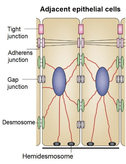 N
N
Not a single diagram that I have seen on desmosomes and intermediate filaments has mentioned the importance of the presence of mitochondria in building and breaking down these structures.