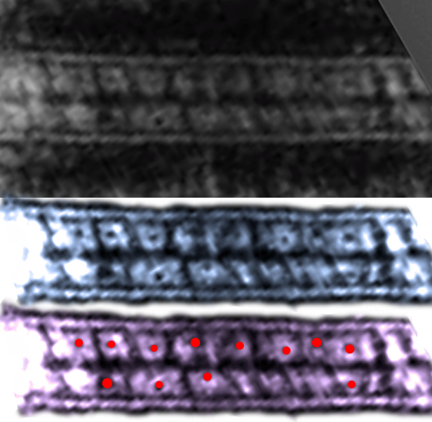
Looking further at the great publication by He, Cowan and Stokes in Science 2003, i just was surprised to see what looked like the most orderly set of dots in the intercellular space on either side of the central dense line (where the desmocollins and desmogleins are supposed to be hooking up in their “velcro-like” attachments. I did a screen print of their micrograph (top image which I did not manipulate)and pasted into photoshop to enhance the pattern with the burn tool (shown in the two bottom images) as an overlay of two separate layers of their original image. The dots and grid are so obvious as to be almost “silly” and if you don’t see what i am talking about…. check out the red dots within the grids in the two images below their micrograph. I have looked at hundreds of desmosomes…. now i have to go back and see whether any of my more opportune images show dots within the grid (which I have seen before. Here the Y and alternating pattern of the intercellular grid is very very apparent.
 It is still possible i think, to have the Y formation be the most prominent, and also repeated in a very orderly fashion (one difference between what i see and what the paper above suggests… when they call the desomosomal cadherins to be in “a knot”. I don’t think disorder is part of this structure… but there rather there is an order which is just a little difficult to detect, owing to the very many angles (at a thickness of 90nm) to bisect a desmosomal spot and untying those possibilities (not to make a bad pun on their title) is not easy. U havn’t yet found a publication that speaks to anything that would make these central densities.
I know there are no mitochondria in their micrgraphs… and that is ultimately what I am curious about, but just in case you havn’t figured it out yet….. biology is totally amazing.