After so many years of looking for articles on this topic (I actually wondered how this phenomenon could go unnoticed for so many decades) i did find one manuscript (which references two others) by Deane et al 1966 which mentions them and shows several micrographs (sometimes pretty low manification). They found these first in fetal and neonatal tissues, so this is a clue and also important for the concept that I have which is that in tissues with higher rates of cell division that also are epithelia which connect at environmental interfaces (meaning the “outside” whether that is physically outside or not) have an active mechanism for building and disassembling desmosomes. They felt it was a one-way passage…. i think it is more a modeling-remodeling phenomenon. So this was nice but bitter sweet at the same time since I thought my early paper in a very obscure journal in the 1980s was the first.
(I am going to use this acronym…desmosomal mitochondrial tethers regardless of how silly it sounds just because it is too time consuming to type out 32 characters when 7 will do, my apologies.)
miller et al, desmosomal mitochodnrial interactions_1985
deane_1966_desmosomal mitochondrial complexes
In figure 1, and enlargement inset called figure 2 (a portion of which is below) of their article, they make a cute comment…. about a desmosome beginning to develop nearby to a reasonably well defined desmite. It would be better perhaps to have recognized that there were two possibilities — one, that it was a tangential cut to a sequentially placed tether along a single mitochondrial outer membrane, and the other that it was a desmite “being built or broken”. This little area of not completely well defined junction they label with a black arrow. You can see that it is not quite a desmosome and the mitochondrion really isn’t that close to it. The label TF (i don’t know why it is in italics) surely stands for tonofilaments…. which I refer to as intermediate filaments.
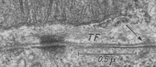
Just using their image and their micron marker (0.5mu = 500nm) it seems that their cut in this desmosome is just off center but close (diameter with the annulus) at about 286nm (central cut yields a desmosome of 300nm) and a diameter of 214nm excluding the annulus. About 162nm span the distance from the outer mitochondrial membrane to the plasmalemma membrane of the desmosome of the cell on the same side. A quick measurement for the thickness of the extracellular space of the desmosome in this image was about 18nm. About 15nm /2 for the dimension of the annulus area on either side of the desmosomal spot. This micrograph shows unequal annulus dimensions. Lovingly I point out the scratch (likely on the old acetate negative) lower left side of the desmosome… and in sympathy say, my negatives and micrographs have many such scratches. One thing to not is that 3-5 intermediate filaments are lying almost parallel to the plasmalemma and the outer mitochondrial membrane… certainly NOT like the diagrams seen routinely — see my post with desmosomal diagrams with hairpin (wrongly directed) lines for intermediate filament attachments to desmoplakin molecules.