Daily Archives: February 26, 2018

Breast cancer and the environment newsletter 6

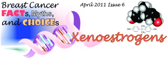
Many xenoestrogens (chemicals which the body is tricked into thinking is estrogen) can pose risk for breast cancer. One such chemical is bisphenol- A, (Figure 1) which can interfere with normal estrogen-dependent functions. The increasing prevalence of xenoestrogens in the environment may partly explain the increasing incidence of breast cancer, though direct evidence is not overwhelming.

The use of bisphenol-A (BPA) in the production of plastics began around 1891 (e.g. in baby bottles, food containers, water main pipes, and laboratory and hospital equipment). Its estrogen-like (in this case, estrogen-disrupting) effects began to be noticed in the 1930s. Prenatal exposure to BPA (in rats and mice) changed mammary tissue and led to cancers in adulthood. Other animal studies used BPA to show that when breast cancer has been induced by a carcinogen that risk was increased further. If the animal studies correlate with humans, then even a small exposure to BPA could cause an increased risk for breast cancer.

Exposing cells in culture to BPA can cause neoplastic transformation of human breast epithelial cells.

“Consumer groups recommend that people wishing to lower their exposure to bisphenol A avoid canned food and polycarbonate plastic containers (which are identified as , unless the packaging indicates the plastic is bisphenol A-free. The National Toxicology Panel recommends avoiding microwaving food in plastic containers, putting plastics in the dishwasher, or using harsh detergents on plastics, to avoid leaching”. (thanks wiki)

Some estimate that 92% of canned goods with plastic liners have BPA. WHEN THE BRAND NAME on your canned soup is “Healthy Choice” and the BPA is 323 ppm, it’s kind of sad. I think its going to be a while before companies get the message…. therefore BUY FRESH
http://organicgrace.com
http://treehugger.com

HERE IS THE PDFPRG-DEH_Issue_6

Mechanotransducer (the desmosome)

Mechanotransduction is known to be an important biological process rendering tissues both resilient and responsive towards their mechanical environment” says the first line of the introduction of this article.”–   mechanotransuction: definition:(biology) Any cell, etc. that generates a measurable response to mechanical stimulation–so this definition includes many many organelles, including the desmosome.

It is fun to think about the desmosomal – mitochondrial tether as the energy supply and the

Adherens junctions and desmosomes

Narrowing of the intercellular space = about 5nm where the cadherins couple

I am not sure why the intercellular space is recorded by What When How as different dimensions (beside the adherens junctions 25nm vs 20nm, the height of the adherens junction itself) and of the intercellular space beside the cardiac desmosome as 35nm and the height of the desmosomal intercellular space as 20-25nm. It seems to me that if one is comparing intercellular space heights, that one really needs to set some parameters and get some comparisons. This could be a massive job, as there are so many variables, not to mention fixation parameters, tangential sections, membrane proteins, cell types and where on the plasmalemma one is going to attempt to measure.
Just in four desmosomes (between hepatocytes from syrian hamster) all fixed the same, similar ages and reasonably good section orientation the following is clear.
1. the intercellular space is pretty variable
2. desmosomes are going to have a slightly smaller intercellular dimension than adjacent intercellular space
3. the center dense line extends beyond the outer desomosomal plaque proteins
4. there is a density in the plasmalemmae as an annulus or ring around the desmosome but intercellular space is wide
5. the separation of leaflets of the desomosomal plasmalemma is just a little more distinct than distant plasmalemma
6. the outer leaflet of the plasmalemma at the desomosoe seems to be quite rigid

See below, four examples and the relative wide range of reduction in intercellular space. Syrian hamster — routine electron microscopy, red dots intercellular space remote from the desomosome, blue dots height of intercellular space at the desmosome. Two of the images – right top and bottom – have double mitochondrial tethers. Left side images have a single mitochondrial tether. Right top and bottom have cross and longitudinal sections of intermediate filaments, respectively adjacent to the mitochondria. In both cases the outer mitochondrial membrane also has a rigid look.