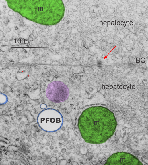
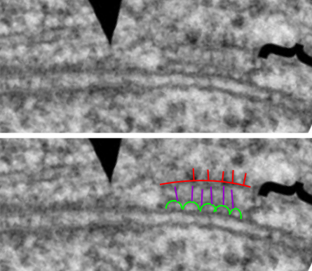
Rabbit liver, two hepatocytes revealed in this electron micrograph. Three possible, one definite, inclusion left over from the infusion of perfluorocarbon, from my notes it looks like perfluorooctylbromide (19.4%PFOB 5%F68), sacrifice dates show that it was more than one year affter infusion. The mitochondria are pseudocolored green, the microbody purplish, the PFOB inclusion complete round (the density of PFOB is greater than water, which I surmise is the reason these structural footprints are completely round and empty) and the very tidy layer of proteins around the perfluorocarbon droplet and bounding membrane are blue. A small (less than 100nm) droplet is typical for the remnants of infusion of the more desirable perfluoochemical blood substitutes. This particular droplet does not show a “black cap” of enzymes which often accompany the droplets. Nice little desmosome between these cells and a bile canaliculus barely seen on the right hand middle. Golgi in the lower cell at bottom with another microbody (peroxisome – which i should have colored as well. My recollection is that infusion of some perfluorocarbons was accompanited by proliferation of peroxisosomess (Neg 5890_block_4783_28000x.
Category Archives: Ultimate order, the cell
Apoptotic nucleus
I wish i had taken a higher mag picture of this apoptotic nucleus as well as identified the cell type. I do know it is from animal #500 in a study of aged ghkaα-/- mice, this particular KO was 12 months old. It could be a parietal cell since in other image parietal cells are shown to have a “glassy protein” (stains without much texture and eosinophilic with H&E) (just recollection here). The RER is greatly, ribosomes are very closely packed (indicating the production of large amounts of possibly large protein products. Checking the fairly electron lucent contents of the RER there doesn’t seem to be any obvious oligomerization, polymerization or layering of the contents. The light areas in the perinuclear space and around is dilated RER. It could also be a zymogen secreting cell.
One nuclear pore on the middle left nuclear membrane, and a part of another to the right and above that one. Packed ribosomes are in array on the RER membranes (the black and white cytoplasm pictured around the nucleus), lots of condensed chromatin (purple) which may be exaggerated by the section being somewhat tangential to the widest diameter of the nucleus, and adjacent euchromatin (gold) which in this case looks pretty dark (not just because i pseudocolored it). There is a prominent perichromatin granule just to the lower right of center (black) and interchromatin group (sometimes called nuclear speckles which to me makes no sense) is light violet. Pinkish spot in the interchromatin granule is probably just euchromatin?
Annulus around desmosome – freeze fracture
Here is an image from a letter to the editor (which i didn’t read thoroughly but was just looking closely at the freeze fracture of tight junction encircling a desmosome) and saw what I had seen in the transmission micrographs of thin sections: that is…. a small ring around the desmosome which i called the annulus which is restrictive and different than the intercellular space nearby. This particular freeze fracture micrograph (original found here) by Yasuo Kitajima shows it clearly, yet is it not marked. I don’t know what the function is but clearly a “no fly zone” surrounds the desmosome. Top micrograph untouched, bottom micrograph with bar marker and blue dotted lines showing part of the annulus. It is kind of nice that this particular desmosome is approximately what the literature suggests in terms of number of nm for the diameter.
so, next question is what are the semiregularly raised areas…?
Weibel-Palade bodies: fun stuff
I have seen these… at the present moment I am looking over the published literature for Weibel Palade bodies (WPD) in order to determine if there is ever a place where ribosomes are present on the bounding membrane. So far, none. This makes this cell adjesion group of molecules, while oligomerized into very beautiful structures, some like tubules, they are quite different from the intracellular granule purported to be a surfactant protein (possible SP-A) found in the cytoplasm of alveolar type II cells in the lungs of some species… which have a very precise localization and number of ribosomes on their limiting membrane (opposite sides of the long dimension of the granule, and approximately 4 ribosomes per 100nm).
Weibel-Palade Bodies (WPB) | Layered granules in alveolar type II cells |
At least 12 different proteins are found in granules | Undetermined number of proteins in type II cell granules |
Recovered as oligomers in clathrin coated vesicles | Recovered, but not recycled or re-internalized as oligomers |
Granules as single membrane bound SMOOTH ER | Granules as single membrane bound with end to end distribution of ribosomes, |
Interacts with cell elements in blood | Interacts with fluid elements in alveolar space |
Endothelial cell product | Epithelial cell product |
Interacts with white cells and platelets | Interacts with alveolar macrophages |
VWP apparently oligomerizes to influence the shame of WPBs | Oligomerization of the SPs involved in intracisternal bodes of alveolar type II cells create the granuel shape |
WPBs maybe storage (albeit a small percent of total) for VWP | Granules in type II cells maybe for surfactant protein storage |
WPBs have P-selectin in small quantitites, i wonder if these are for binding of clathrin proteins to initiate recycling from the secretion bubble structures after exocytosis of VWP? | Re-uptake, recycling of surfactant proteins is not so obvious (but happens) not big obvious vesicles… at least not that I have seen. |
The cell: a polymer chemists playground
I think it is just overwhelming to see the order “of life”. Starting out with a search for patterns in electron microscopy for desmosomes has led me to find cell adhesion molecules a really fascinating, perhaps the ultimate, cell-environment interface. So I will go on a hunt, just for the pure joy of finding them.
Von Willebrand Factor (article) bar=10nm. “This factor serves as a docking station or molecular bus to which numerous proteins bind during the formation of blood clots.” (quote from article linked above by Zongchao Jia).
NICE Review of a few cell adhesion molecules
It is worth reading this nice review article which deals with adherens junctions and also some other cell adhesion molecules.
Oda and Takeichi 2011 have this nice electron micrograph or cell-cell adhesion, but it is even more interesting to me that the area described with the confines of their arrows, and the area within their bracket, does not show as distinct an organization as the area inbetween their designated zones… Haha. see bottom two micrographs, their photo unretouched, and the area where I see amazing order, an area which is very organized.
semicircular arrangement (black lines), likely proteins which attach to whatever membrane proteins the circles are, and then the red lines as some molecule which links the former with some cytoskeletal element. There is a great microtubule over by their arrows, i don’t know what is under their bracket.
Just a note: it looks like the primitive junctional complexes in drosophila are the result of one-size-fits-all type molecule with little segments snipped out as DE-cadherin has every thing but the kitchen sink there….including laminin, proteolytic, and EGF-like domains.
Natural inspiration for bargello
Right hand:left hand
Sometimes I get dismayed at what the university of cincinnati (along with cincinnati children’s hospital) finds acceptable. Just one parking lot away from the department of environmental health, where government has funded the removal of lead and asbestos in an effort to make the city a safer place to live, are two giant yellow excavators pulling down 100 year old neighboring houses (not that i am bemoaning the loss of less well kept property) unquestionably filled with lead paint, asbestos, mold and piles of trash billowing up dust clouds that would signal that much is airborne that should NOT be airborne. So cincinnati children’s is (has/continues) expanding into a very old neighborhood adjacent to it) but doing it in a way that is environmentally reckless (in my opinion).
I have met and talked to residents in those homes… I remember one in particular at the south-east corner of burnet and erkenbrecher who was a long last holdout before he sold to children’s hospital. I have spoken with the owners of two of the very lovely old brick victorian houses on erkenbrecher, one a professor here, and i wonder whether greed will overtake historical value for them as well.
two issues here: 1) environmental health is important — and it is the biggest department at the university of cincinnati BUT why can its neighbor (also part of UC) disregard the information, persuasion, good policy of its back-door neighbor and pollute the environment of its own “house” so to speak. I dont get it. Doesnt the right hand know what the left hand is doing. 2) second issue is just preservation of history.
CAM: apology about cell adhesion molecules – I wish i could find what i am looking for
It is overwhelming to move into a new field of study. In this case one borne out of looking at desmosomes. I am starting at the beginning with this overview for myself primarily, if someone else finds it useful, so much the better.
Cell adhesion molecules — a mind-blowing statement about how the cell has learned to interact with its environment over the last 3.9 billion years. It should not be surprising that a huge number (hundreds if not thousands) of molecules are out there in the cell membrane touching and feeling what is around…. either another cell, or matrix or the extracellular milieu. The adhesion molecules in desmosomes are part of this elaborate system to “group” “assemble” and become an “organism”… present in insects in highly organized and organized junctions. So here is a very basic summary of the Cell adhesion molecules.. a basic diagram without complete listings but helpful to me. There is a short list of some representatives of the four superfamilies on the left of the graph, no attempt made to be inclusive, but more to show diversity.
Think of the cell as a tiny tiny pincushion, the cell membrane packed with proteins which have an intracellular domain, an intracellular domain, and an extracellular portion, each little molecule protruding like an feeler into the environment — so to speak– to sense portions of, to signal to, to interact with, and/or adhere to. I marvel indeed.
A little acronmistic thing here…. of the four superfamilies of cell adhesion molecules it is kind of nice that two of them, the Cadherins (though i did find a report that says some cadherins lack a calcium binding domain…. maybe these are mutated..according to the author?) and the C-type lectins both are calcium dependent and begin with the letter C, while the other two begin with the letter I – and are calcium independent — that is Integrins and Immunoglobulins.
One other kind of cool thing is that two of the C-lectins, one in particular (that is surfactant protein A) can oligomerize into wonderful patterns seen electron microscopically, and the cadherins, desmoglein and desmocollin, appear to have a very organized extracellular and intracellular status as well.
Dental impression alginate
This is just a quick post and may not be relevant to anyone in the cosmos but I had several TEMs of dental alginate (in this case it was Kromopan) that were made when I was trying to figure out a way to quanatify gingival overgrowth in mice. I am not sure that a way was found but we tested several alginates to see which one gave the best tooth “footprint”, gum, and bone delineation. This was just a fun gemish. I dont know if i will spend the time to look up the structure and compare what i saw to what is known about the alginate. But this is an artsie-craftsie version of the original TEM (lower right). Fixed in buffered aldehydes three days after molding in mouse jaw, and routine processing for TEM. Bar marker=1 micron.