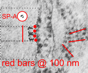
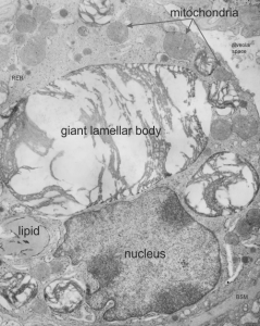
Cat (feline) alveolar type II cell RER: is this a layered granule in the RER? It has only about two of the characteristics of what I think is surfactant protein in the RER which is three species makes a highly layered secretion granule which fits a little nicely with a parallel arrangement of a four stack of surfactant protein A. (that is probably not the best way to describe it, but a vertical stack of two mirrored surfactant protein A groups, stacked again and then replicated side by side many times seems to fit what is seen in some granules). Rabbit alveolar type II cells have some indication that a similar RER granule might be present, other species, not so much. Hamster and cat come close, but do not really fit well. BUT case in point, here is an alveolar type II cell from a cat, which shows RER profiles with one of the important criteria for being called a surfactant protein granule, and that is the loss of ribosomes on the long axis of the granule (typically parallel to the layering) and the presence of stacked ribosomes (seemingly 4 per period (one period being two outer dense layers and one central pretty much dense layer), one ribosome on the dense bands each, and two ribosomes on either side of the central band)…. so here just on the end of this profile of RER in the cat alveolar type II cell is a stack of 4 ribosomes, and then tangential off to the right is a series of 4 ribosomes stacked as well. Image below and inset are not enhanced or photoshopped except to increase contrast slightly. dust and scratches remain. bar = approximately 100 nm (determined from the presumed 27 nm (give or take) mammalian ribosome size.
Category Archives: Layered intracisternal protein granules in mammalian lung
Rabbit alveolar type II cell: criteria met for protein granule?
I revisited the protein acumulation in the alveolar type II cell of a rabbit (this particular rabbit was 79 days post infusion of an emulsion of oxydecalin 10% and F68 25% at 60cc/kg intravenously, on 9 9 1987. negative 11320, block 31750, and this is one of only two locations where there was a stacked (or single) profile of RER which had some of the characteristics of an intracisternal protein that might be made up of surfactant protein, in particular surfactant protein A. The criteria met include: 1) stiffness of the portion of the RER which has the protein inclusion visible, 2) loss of ribosomes on the longitudinal axes, but ribosomes on the growing end(s), 3) some evidence of layering within the granule (see inset where i highlighted what i think is evidence of layering) 4) a parallel stacking. I have considered this an equivocal demonstration of a surfactant protein granule in the alveolar type II cell, since the layering is really not that prominent. The bigger issue is that, if one considers the ribosome of the rabbit, to be similar in size to other mammalian ribosomes (I have taken as an average about 27 nm), then the width of the profile at its narrowest dimension is about 1.5 times wider than the layered granules seen in guinea pig, ferret and dog. So this is an issue. See for yourself in the image below. (two prominent scratches stamped out using photoshop, contrast increased also, otherwise no processing). Red dots=ribosomes, dotted boxes show areas of inset. Insets show areas of possible banding which I highlighted with the burn tool in photoshop. So original on right, emphasized on left.

While you are on this page, check out the awesome glycocalyx on the apical plasmalemma folds and projections in the upper right corner of the electron micrograph.
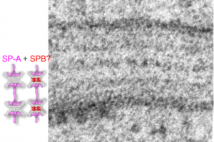
4 ribosomes – four SP-A molecules?
Another view of an image of an RER granule from an alveolar type II cell. where an actual SP-A shadow cast molecule (from the internet) was duplicated, mirrored vertically, both copied again, and pasted so that four molecules are stacked. These then were placed next to an opportune profile of an RER granule which clearly showes 4 ribosomes in a period…. As previously mentioned, one ribosome for each of the most dense layers (that is at the widths of the single periods) and two ribosomes within the period, one on either side of the central dense band. 4 ribosomes per period in all…. which pretty much matches the concept of four SP-A molecules end on end as imaged here. Still thinking on this, comments (to my personal email, not on this blog page) are welcome.
SP-A and SP-B together in the alveolar type II cell granules?
Does the vitro replica of surfactant speak to SP-A and B participation in the alveolar type II cell granules?
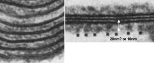
So do you think that SP-A is the main protein component of the alveolar type II cell granule? SP-B has been shown to work with SP-A in several circumstances and particularly in forming tubular myelin lattice structures. SP-B is a pretty small and flat molecule (but not as flat as DP-C, and it diagrams seem to place it wort of “surface-wise) to the plasmalemma bilayer. and if i remember right, maybe the bilayer elements of the alveolar space. Here is a cut and paste from a paper from the “before times” by Williams et al, 1991, where they show artifically created showing some lamellar ultrastructural similarities with the two dense lines and central punctate line seen in RER granules of alveolar type II cells. (but I am not sure about size) thought the mention of 20 nm is there, because i don’t know if that is a measurement from one dense lipid layer to the center punctate line, or all the way across the lamellae. Her reference is HERE. and a cropped figure from her publication is below. The match is certainly not perfect, but suggestive of possible structural organization of SP-A and lipid.
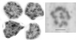
Comparison between shadowed SP-A and tangential figures in electron micrographs
Voss et al, JMB 201 : 1988 published shadow cast images of SP-A (which I did a screen print on (right hand box with bar marker of 20nm for all images) and images from my own micrographs of the layered RER granule (intracisternal body) in alveolar type II cells found in some species. The bar markers are just a little different, which is not at all surprising, but the figures themselves and general size matches are pretty striking. I have compared with structures derived from tangential sections of RER granules (intracisternal bodies) in alveolar type II cells (4 samples on the left).
Alveolar type II cell in rat: vinyl chloride and vitamin C
I am posting this not really as a huge project, but just as a reminder to self, and to those interested that sometimes data are lost and forgotten but can still have meaning. The study mentioned here was just part of a very large study which was conducted by a good friend (late, actually many years passed on) who was very careful in what she did, and didn’t press conclusions or publications beyond their real findings. She was also a good friend. Dr. Martha Radike (who did these inhalation experiments at the University of Cincinnati, College of Medicine, Department of Environmental Health, back in the 1980s. The test animals (160 male Hartley guinea pigs) represented significant effort. I did some of the electron microscopy but never completed a manuscript on the animals (and they represented only 18 out of the sum total of the experiment.
My interest in the guinea pigs was revitalized while doing a bigger review of over 1400 micrographs looking specifically for a granule of surfactant protein in alveolar type II cells. So this study I brought up out of the archives for that purpose and found a summary, which I have posted HERE as a pdf for anyone who is interested. vinyl_chloride_vit_C_guinea_pig_lung
Here is an enlarged image from that pdf.
Not much question that these are the same structures in type II cells
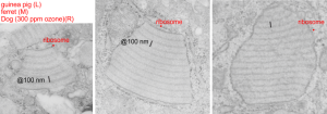
Electron micrographs of portions of alveolar type II cells: Two micrographs on the left are from my research, guinea pig on the left, ferret in the middle. The image on the right comes from a publication in 1973 by Stephens, Freeman, Stara and Coffin which had to do with exposure of beagle dogs to ozone. The claim that ozone increased these protein bodies (which for all intent and purposes match the protein body granules that I have seen in my animals), which also increased with age, particularly. I attribute these to some kind of immune response to environemetal exposure, and/or immune disturbance (as in the case of the guinea pigs, maybe an entire room of guinea pigs in the experiment were infected with some agent that increased the proteins in lung for innate immune response. Regardless, the point of this image is to demonstrate electron micrographs from these three species, of alveolar type II cells, showing what stress can produce in the way of organellar responses (if one can call this granule, an organelle — which begs the issue of calling Birbeck granules in Langerhans cells… organelles–again in which case these might be SP-A granules haha). I have marked the approximate 100 nm bar in all three images, and made a sample ribosome size in each as well for suggesting magnification. The periodicity found in the RER of the beagle electron micrograph I think was stated to be less than 100 nm… (76 nm or something instead).. you can choose. What I have called 100 nm is nominal.
Text book pictures of untreated and ozone exposed guinea pig alveolar type II cells
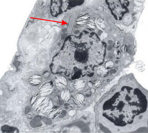
There is a book called Diagnostic Ultrastructural Pathology – A text atlas of case studies. Volume 1 ( Ann M. Dvorak, Rita A. Monahan-Earley) which on page 218 posts figure 182 (reproduced here – I thank them). It shows clearly that, while they were interested in lamellar body ultrastructure before and after ozone exposure, you can be sure that the most important structure for me is the “cisternal body” that flattened and layered protein structure that I see in the upper right-ish corner of their micrograph. Guinea pig must have some very robust mechamism for responding to environment, producing proteins (presumably some surfactant proteins) and in this case, after ozone they do not seem to become more prominent (according to this chapter in the book) as they do in dog (see previous post), but actually become less conspicuous.
Dimensions and position within the cell are classic “cisternal body” which I really wish I could rename SP-granule. Red arrow points to the RER granule.
Eureka, “ah ha” !! I found reference to a study on dog lung which shows alveolar type II cell granules (cisternal bodies).
There was a group of scientists in california (I could not remember which research institute) that kindly sent me some plastic-embedded tissues of dog lung, since I had found a paper someone there that described layered protein granules in alveolar type II cells like I had seen in ferret and guinea pig and had ask them for some tissue. I could not (after 35 years, remember their names). HERE IT IS! awesome, though at least one of the researchers is deceased (J. Stara). I am taking some of their electron micrographs, cropping, editing contrast (only) and placing them next to the ones I found in guinea pig and ferret. Perfect matches. This publication is so old (1973) that there is no way that surfactant proteins could have been implicated as possible contributing factors to these granules that appeared in their ozone treated beagle dogs. The surfactant proteins had not yet been described. Ozone caused what was described (for lack of background information on overall type II cell function in those days) “metabolic alteration ….. this toxic agent”, which of course is a statement which NOW in 2016 in light of decades of research on surfactant and lung function looks patronizing.
Their RER measurements made this “approximately 754 A (75 nm) is not that far off from what was calculated by my own measurements 100nm (microscopes differ in magnification tables, and so neither is likely to be exact).
Because of their reporting of small, sometimes stacked “bar like” structures such as I have seen in untreated dogs (and looking a lot like what I have seen in rabbit type II cells). it is likely that an “excessive” amount of surfactant protein is produced and maybe stored in a multi-meric configuration) within the RER awaiting utilization in assembly within lamellar bodies or multivesicular structures. Stevens et al, 1973, mention (yippie) that there is a dense line which runs parallel to the length of the RER in these infrequently seen structures.
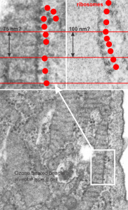
Ribosomes in the beagle exposed to ozone that border the RER profiles on the growing ends of the layered protein do look to be in similar positions with regard to banding of the granule to those found in ferret. My inclination is to think that there is a ribosomes on either side of the central band in each period (beagle on the left, ferret on the right – where central band is not really visible in this view) and a ribosome at the point of the darkest band (counted on two sides if there is only one period but counted only one one side if there are multiple periods) making each periodicity have three ribosomes, not counting the dark band of the adjacent period. One on a dark outside band, two on either side of the inner lighter central band. In the figure below, see comparative diagrams. the red lines with black arrows and text indicate what they think is 75 nm and I think is 100 nm (right) (give or take). Ribosomes are red, distance arrows are black, inset is enlarged from white box on bottom electron micrograph, dog on left (from Stephens et al, 1973, top image of figure 3, used without permission), ferret on right, my studies. Ribosomes are the bar marker (25-30 nm each). It is important to note that not all RER cisternae in their exposed animals showed periodicity…. just like those in guinea pigs… which I attribute to tangential sectioning and multiple intersections of different “growing ends”, and also the sudden transition from flattened cisternae of RER to the layered granule.

