Category Archives: Electron microgaphs of lung

Various species of mammal and maybe a non mammal now and then have been obtained and examine using routine transmission electron microscopy. These are summary images.

Rabbit alveolar type II cell: criteria met for protein granule?

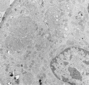
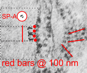
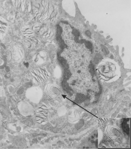
I revisited the protein acumulation in the alveolar type II cell of a rabbit (this particular rabbit was 79 days post infusion of an emulsion of oxydecalin 10% and F68 25% at 60cc/kg intravenously, on 9 9 1987.  negative 11320, block 31750, and this is one of only two locations where there was a stacked (or single) profile of RER which had some of the characteristics of an intracisternal protein that might be made up of surfactant protein, in particular surfactant protein A.  The criteria met include: 1) stiffness of the portion of the RER which has the protein inclusion visible, 2) loss of ribosomes on the longitudinal axes, but ribosomes on the growing end(s), 3) some evidence of layering within the granule (see inset where i highlighted what i think is evidence of layering) 4) a parallel stacking. I have considered this an equivocal demonstration of a surfactant protein granule in the alveolar type II cell, since the layering is really not that prominent.  The bigger issue is that, if one considers the ribosome of the rabbit, to be similar in size to other mammalian ribosomes (I have taken as an average about 27 nm), then the width of the profile at its narrowest dimension is about 1.5 times wider than the layered granules seen in guinea pig, ferret and dog.  So this is an issue.  See for yourself in the image below. (two prominent scratches stamped out using photoshop, contrast increased also, otherwise no processing). Red dots=ribosomes, dotted boxes show areas of inset. Insets show areas of possible banding which I highlighted with the burn tool in photoshop.  So original on right, emphasized on left.

rabbit_alveolar_type_II_granule_SP-A
While you are on this page, check out the awesome glycocalyx on the apical plasmalemma folds and projections in the upper right corner of the electron micrograph.

Hamster: alveolar type II cell

Another example of a species in which I have hunted from layered granules in the alveolar type II cells is hamster (though only a few animals have been studies). There are, as in cat and rabbit) frequent examples of RER which has a slightly dilated appearance, and also has obvious protein density within the lumen of the profiles, but lacks all but the very faintest appearance of layering, and also does not abide by the criterion for layered granules in that ribosomes stud all aspects of these RER profiles, and the rigid looking, ribosome free, part of the granules seen in ferret, guinea pig and dog, thus one has to assume either that surfactant protein A is sufficiently unique in the three species above where granules can organized into a regular substructure, or that something else altogether is responsible.  hamster, male, animal# 3, neg 7278, block 24733,

hamster_lung_type_II_cell

Cat lung: alveolar type II cell

Cat lung: alveolar type II cell, with arrows to three profiles of endoplasmic reticulum which have a structure not typically found in alveolar type II cells of common laboratory species (mouse, rat). These are rounded and it is not actually that hard to see some striations in them, particularly the one on the most left.  In a series of micrographs from untreated cats (taken incidentally from other studies) no sturctures with highly visible layering in the RER were seen, which doesn’t mean these profiles are not surfactant proteins, it just means that they are not organized as seen in ferret guinea pig and dog alveolar type Ii cells in other studies. negative 7268, 24564, cat #3, untreated (gender?, age?)

cat_lung_type_II_cell

Rabbit lung: post intratracheal instillation of iron oxide and dibenzo cabazole

Rabbit lung: post intratracheal instillation of iron oxide and dibenzo cabazole, electron micrograph. This is not a great picture, but i post it just so one can appreciate that there an area of odd membrane swirling in the middle left, and a small area just like it in lower left. The cell has a lobular nucleus and there are iron oxide (probably with DBA alongside) within this cell. I don’t think it is an alveolar parenchymal cell, but likely an inflammatory cell. A type II cells from the same microgrpah has lamellar bodies, which this cell does not, which further indicates it as a migratory cell. Animals given DBC do get tumors. neg 4447, block 17793, Rabbit #32-3.  Sorry the micrograph has stain ppt on it, but it is worth looking at just for the layered structure.

rabbit_lung_iron_oxide_dibenzo_carbazole


4 ribosomes – four SP-A molecules?

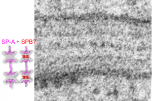
Another view of an image of an RER granule from an alveolar type II cell. where an actual SP-A shadow cast molecule (from the internet) was duplicated, mirrored vertically, both copied again, and pasted so that four molecules are stacked. These then were placed next to an opportune profile of an RER granule which clearly showes 4 ribosomes in a period….  As previously mentioned, one ribosome for each of the most dense layers (that is at the widths of the single periods) and two ribosomes within the period, one on either side of the central dense band.  4 ribosomes per period in all….  which pretty much matches the concept of four SP-A molecules end on end as imaged here. Still thinking on this, comments (to my personal email, not on this blog page) are welcome.

ribosomes_SP-A-4_and_4

Does the vitro replica of surfactant speak to SP-A and B participation in the alveolar type II cell granules?

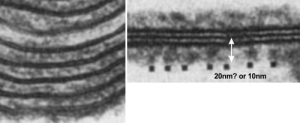
So do you think that SP-A is the main protein component of the alveolar type II cell granule? SP-B has been shown to work with SP-A in several circumstances and particularly in forming tubular myelin lattice structures. SP-B is a pretty small and flat molecule (but not as flat as DP-C, and it diagrams seem to place it wort of “surface-wise) to the plasmalemma bilayer. and if i remember right, maybe the bilayer elements of the alveolar space. Here is a cut and paste from a paper from the “before times” by Williams et al, 1991, where they show artifically created showing some lamellar ultrastructural similarities with the two dense lines and central punctate line seen in RER granules of alveolar type II cells. (but I am not sure about size) thought the mention of 20 nm is there, because i don’t know if that is a measurement from one dense lipid layer to the center punctate line, or all the way across the lamellae. Her reference is HERE.  and a cropped figure from her publication is below. The match is certainly not perfect, but suggestive of possible structural organization of SP-A and lipid.

in_vitro_surfactant-lipids_and_SP-A

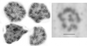
Comparison between shadowed SP-A and tangential figures in electron micrographs

Voss et al, JMB 201 :  1988 published shadow cast images of SP-A (which I did a screen print on (right hand box with bar marker of 20nm for all images) and images from my own micrographs of the layered RER granule (intracisternal body) in alveolar type II cells found in some species.  The bar markers are just a little different, which is not at all surprising, but the figures themselves and general size matches are pretty striking. I have compared with structures derived from tangential sections of RER granules (intracisternal bodies) in alveolar type II cells (4 samples on the left).

TEM_and_shadowed_SP_A

Guinea pig (or ferret) alveolar type II cell lamellar body: projection core?

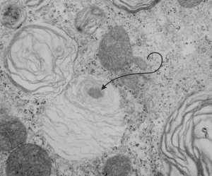
guinea_pig_lamellar_body_projection_coreIn lung, the type II cell has lamellar bodies, the secretion granule of surfactant, but not all species show a projection core within those lamellar bodies. I don’t know if guinea pig has ever been described as having projection cores in their lamellar bodies, but this looks either like a portion of a multivesicular body-fusion with lamellar body, or maybe a projection core (which should probably have more layered substructure).  It has a different electron microscopic appearance than lipid does, and lipid droplets, seen as mostly textureless round areas, are found in lamellar bodies very frequently, and in many species in my experience.  Here is a crop from a type II cell from guinea pig lung and the lamellar body (end of pointer) seems to me to have a projection granule. (6481_M8005 guinea pig #51, vinyl chloride exposed plus 2mg vitamin C you can access this pdf with specifics and details of animal exposures vinyl_chloride_vit_C_guinea_pig_lung )

I also went quickly through the electron micrographs I have for dog lung (467 lamellar bodies quickly counted, with 24 lamellar bodies equivocal for a projection core, but not really a good match, looking much more like areas where there is multivesicular body material.

Also, perusing over 1000 lamellar bodies in electron micrographs from ferret my conclusion is that there are many little lipid droplets within lamellar bodies, but only a rare protein-like body that might under ideal fixation circumstances be seen as a projection body but still without substructure that is layered (which Ochs 2009 mentions is the hallmark for projection cores. He also states that they do not appear in rodents).  There are many many examples of multivesicular type densities within lamellar bodies though, clearly defined and usually around the periphery of the lamellar bodies. That said, I understand that “fixation” is everything.

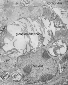
Here is an electron micrograph of ferret alveolar type II cell which has possibly as good an indication from archived photos as any that maybe in some ideal conditions, ferret might actually have some lamellar bodies with some type of density, maybe part of a multivesicular body-core together.  I am not banking on it, but offer this, as one in a thousand example of a lamellar body that might have a projection core. As with guinea pig and dog, most of the roundish things at one or more poles or periphery of a lamellar body look more like lipid than layered protein. Lipid is common.  This could just as easily be a tangential section of the bottom side of a multivesicular body fusing with a lamellar body. And only specific studies will determine which is which, and most likely will turn out to be something of both together.

ferret_lamellar_body_projection_core


Alveolar type II cell in rat: vinyl chloride and vitamin C

I am posting this not really as a huge project, but just as a reminder to self, and to those interested that sometimes data are lost and forgotten but can still have meaning. The study mentioned here was just part of a very large study which was conducted by a good friend (late, actually many years passed on) who was very careful in what she did, and didn’t press conclusions or publications beyond their real findings. She was also a good friend.  Dr. Martha Radike (who did these inhalation experiments at the University of Cincinnati, College of Medicine, Department of Environmental Health, back in the 1980s.  The test animals  (160 male Hartley guinea pigs) represented significant effort.  I did some of the electron microscopy but never completed a manuscript on the animals (and they represented only 18 out of the sum total of the experiment.

My interest in the guinea pigs was revitalized while doing a bigger review of over 1400 micrographs looking specifically for a granule of surfactant protein in alveolar type II cells.  So this study I brought up out of the archives for that purpose and found a summary, which I have posted HERE as a pdf for anyone who is interested.  vinyl_chloride_vit_C_guinea_pig_lung

Here is an enlarged image from that pdf.

vinyl_chloride_vit_C_guinea_pig_lung