Daily Archives: May 3, 2017

Just a quick comment on nuclear organization

It has been my opinion (based on looking at thousands and thousands of slides and micrographs) that the when the cell undergoes apoptosis or is overly stressed, it shows an amazing nuclear bilateral symmetry.  DIVIDED IN HALF (how does this surprise anyone-yet why can I not find this in the literature)– that is to say that that domains in the nucleus which are beginning to assemble, and/or disassemble in response to the cell death signals, such as the interchromatin granule clusters, and the huge nucleolar structures (fibrillar centers and dense fibrillar components) just seem to appear in light micrographs (where sections are relatively thick compared to those for TEM) in a mirrored symmetry.

I have seen this same phenomenon in white blood cells (particularly bone marrow smears) where the granulocytes were stressed and micronuclei, or bar-like bodies, or segmentation are increased by (in some cases in bone marrow smears from Cyp1A1, Cyp1B1 and Cyp1A2 ko mice and their many variations and exposure toxins like benzo(a)pyrene or TCDD), and in other cases such (in vitro) where RNAi knockdown of the highly conserved anti-apoptotic gene, C9orf82, induced apoptosis and cells showed mirrored nuclear symmetry, and in 14CoS cells that underwent massive synchronized hepatocyte apoptosis.
This is part of the symmetry of the nucleus currently being explored with data mining, fluorescent antibody imaging, TEM-CT, and yes, even the old school “eyeball” approach. Here is an image and diagram of one bone marrow smear where bilateral symmetry of a white cell is very obvious, and though the image is almost a decade old, and I queried the literature at the time I could find no publications on domains,  territories, or mirrored images in apoptotic cells. (BTW the cells below in a bone marrow prep are NOT apoptotic but nicely segmented).
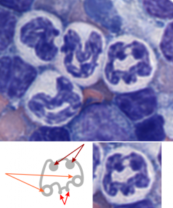
light micrograph bone marrow smear white cell nuclear bilateral symmetry

More microDNAs for cover submission

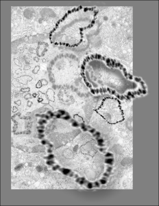
I must have really made a lot of variations for this cover submission, here is another that I modified this morning.  It is a little confusing, but the cell behind is a has a large nucleus (left middle of the micrograph), which is in distress, with some indications of impending apoptosis, mainly the finely granular chromatin at the inner nuclear membrane, and the prominence of interchromatin granule clusters, and a rather large nucleolus (with prominent fibrillar centers and dense fibrillar components (in preparation for the trip down the apoptotic pathway). And on that image I superimposed a dozen or more of the images of microDNAs that Dr. J.D. Griffith gave me to organize into a cover. So the microDNAs, (of which three are enlarged bursting out in dimensional relief) that are mostly black and grey high contrast, while there are many others lesser in size that are superimposed in relief over the original transmission electron micrograph of the cell.

Electron micrograph of nucleus and overlay with microDNAs

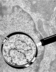
Still working on a cover submission with Dr. J.D. Griffiths microDNA images, superimposed over one of my own electron micrographs of a cell with prominent nucleus and highlighted within that as an inset is an enlargement, over which is superimposed his micrographs of microDNA.

I don’t think this image was selected for submission, but it is pretty nice none-the-less. I am prompted to post these, after looking back over the archives (for other reasons).

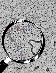
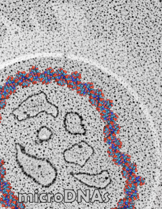
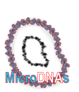
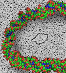
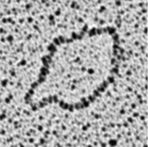
DNA and microDNAs

See these images which were submitted, but not accepted for cover illustrations.  Electron microscopy by Dr. J. D. Griffith, and rendition by Dr. Marian L. Miller.  We were working on a cover submission, and those examples below are just a few that were organized, plus the one I liked best shown in a previous post on this  blog here .  The bottom right image I find this morning while googling, skimmed, and referencing some keyword advertizing company…. this makes me furious. The pasted up DNA is middle right, and all these are Dr. Jack D. Griffiths micrographs of microDNAs.  Shame on you skimmers and spammers and hackers.