Monthly Archives: June 2017

What is this association about

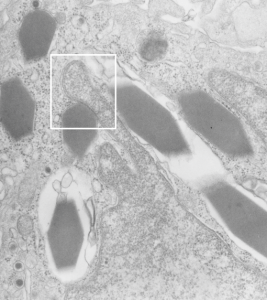
Looking at an alveolar macrophage from an aged guinea pig without treatment (a control) I noticed a cell which contained some very nice crystals, but noticed in particular that when a particular crystal was juxtaposed to the nucleus, that some kind of “order” appeared, and an increase in density of the nuclear membrane was evident.  This would be an interesting thing to research if the macrophages with these inclusions were frequent. At best, I have seen only a few.  As usual, negative 9218_block17082_gpig_301_lung, likely alveolar macrophage, data given just in case someone out there wants backup.

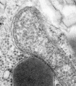
White box is enlarged and contrast enhanced below this image.  Nucleus is a finger like projection from the lower right hand side, and at least 7 crystals are seen.

electron micrograph, alveolar macrophage rhomboid crystals nuclear membrane guinea pig I can visualize these little A-frame things with heads on them about 10 of them on the inner nuclear membrane.  Outer nuclear membrane and granule membrane is really indistinct… owing to the ribosomes in the limiting membrane of the crystal, one could even assume that this crystal might be in the perinuclear space.

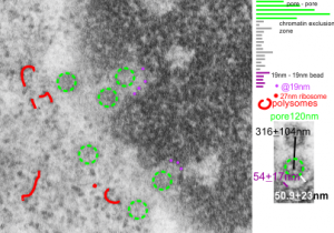
Nuclear pore measurements: pore to pore, chromatin exclusion zone, granule to granule

Three measurements taken in a series. Pore to pore, pore to to edge of chromatin (which i found is called the chromatin exclusion zone, so am sticking with that since it clearly is an active event, and may vary with cell type and cell activity) and inter-@19 nm “beads” that are seen as the organized chromatin just adjacent to the nuclear pore (the first thing seen after the chromatin exclusion zone).  This micrograph is from the same images as a previous post, which is 16027_65718_14Cos_ko 24hr no NTBC measurements.-2a. n=5,  pore to pore=316nm+104nm; n=15, chromatin exclusion zone, 50.9+23nm; n=5, 19nm to 10nm chromatin granule, 54nm+17.7nm

66 names for the devil

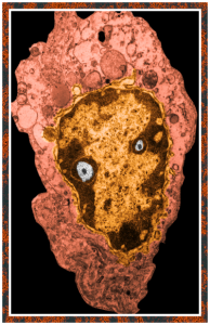
This post is just for fun, as when i was looking at some electron micrographs of the Gclc mouse liver study I found this critter, just with big eyes (one of which i added the pupil to using photoshop — my apologies) but otherwise not retouched.  The exact data are neg17894_block74138_animal706_wcii_NAC. Initially I was interested in the nucleoli, but naggingly amazed about the “devil” appearance of the nucleus as a whole.
Color was of course added using photoshop, all else remains the same. When considering what to call this character i googled “names for satan” and behold, the list of about 66 names from various cultures and groups showed up (cut and pasted from Wikipedia (thank you wikipedia) (below).
Abaddon—(Hebrew) the destroyer
Adramalech—Samarian devil
Ahpuch—Mayan devil
Ahriman—Mazdean devil
Amon—Egyptian ram-headed god of life and reproduction
Apollyon—Greek synonym for Satan, the arch fiend
Asmodeus—Hebrew devil of sensuality and luxury, originally “creature of judgment”
Astaroth—Phoenician goddess of lasciviousness, equivalent of Babylonian Ishtar
Azazel—(Hebrew) taught man to make weapons of war, introduced cosmetics
Baalberith—Canaanite Lord of the covenant who was later made a devil
Balaam—Hebrew devil of avarice and greed
Baphomet—worshipped by the Templars as symbolic of Satan
Bast—Egyptian goddess of pleasure represented by the cat
Beelzebub—(Hebrew) Lord of the Flies, taken from symbolism of the scarab
Behemoth—Hebrew personification of Satan in the form of an elephant
Beherit—Syriac name for Satan
Bilé—Celtic god of Hell
Chemosh—National god of Moabites, later a devil
Cimeries—Rides a black horse and rules Africa
Coyote—American Indian devil
Dagon—Philistine avenging devil of the sea
Damballa—Haitian Vodou serpent god
Demogorgon—Greek name of the devil, it is said should not be known to mortals
Diabolus—(Greek) “flowing downwards”
Dracula—Romanian name for devil
Emma-O—Japanese ruler of Hell
Euronymous—Greek Prince of Death
Fenriz—son of Loki, depicted as a wolf
Gorgo—dim. of Demogorgon, Greek name of the devil
Haborym—Hebrew synonym for Satan
Hecate—Greek goddess of the underworld and witchcraft
Ishtar—Babylonian goddess of fertility
Kali—(Hindu) daughter of Shiva, high priestess of the Thuggees
Lilith—Hebrew female devil, Adam’s first wife who taught him the ropes
Loki—Teutonic devil
Mammon—Aramaic god of wealth and profit
Mania—Etruscan goddess of Hell
Mantus—Etruscan god of Hell
Marduk—god of the city of Babylon
Mastema—Hebrew synonym for Satan
Melek Taus—Yezidi devil
Mephistopheles—(Greek) he who shuns the light, q. v. Faust (Greek)
Metztli—Aztec goddess of the night
Mictian—Aztec god of death
Midgard—son of Loki, depicted as a serpent
Milcom—Ammonite devil
Moloch—Phoenician and Canaanite devil
Mormo—(Greek) King of the Ghouls, consort of Hecate
Naamah—Hebrew female devil of seduction
Nergal—Babylonian god of Hades
Nihasa—American Indian devil
Nija—Polish god of the underworld
O-Yama—Japanese name for Satan
Pan—Greek god of lust, later relegated to devildom
Pluto—Greek god of the underworld
Proserpine—Greek queen of the underworld
Pwcca—Welsh name for Satan
Rimmon—Syrian devil worshipped at Damascus
Sabazios—Phrygian origin, identified with Dionysos, snake worship
Sammael—(Hebrew) “venom of God”
Samnu—Central Asian devil
Sedit—American Indian devil
Sekhmet—Egyptian goddess of vengeance
Set—Egyptian devil
Shaitan—Arabic name for Satan
Shiva—Hindu the destroyer
Supay—Inca god of the underworld
T’an-mo—Chinese counterpart to the devil, covetousness, desire
Tchort—Russian name for Satan, “black god”
Tezcatlipoca—Aztec god of Hell
Thamuz—Sumerian god who later was relegated to devildom
Thoth—Egyptian god of magic
Tunrida—Scandinavian female devil
Typhon—Greek personification of Satan
Yaotzin—Aztec god of Hell
Yen-lo-Wang—Chinese ruler of Hell
More nuclear pore measurements

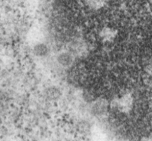
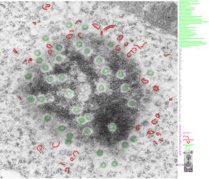
This hepatocyte was from a mouse (#5) which was a 14CoS nujll that did not receive NTBC as a rescue drug. This is 24 hours into the neonatal period.  Nuclear pores have fewer central densities (presumably being proteins transported). I am measuring for distance beside the nuclear pore that the chromatin exclusion zone hoping that differences will show up with various conditions in previous experimental models.

Top micrograph, unretouched, bottom micrograph, pores used in calculation have circles, red dot = a 27nm ribosome, purple dots are areas of the chromatin just adjacent to the chromatin exclusion zone surrounding the nuclear pore.

meaurements of the chromatin exclusion zone around the nuclear pore transmission electron microscopy

Nuclear pores and polysomes and other things

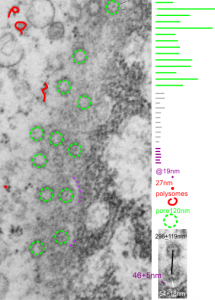
Continuing an evaluation of nuclear pores in various experiments from the past, here is a microgaph from 17709_65053_14CoS-/- NTBC 1yr. I used some of the opportunely sectioned “almost top down” cuts of nuclear pores to look at the inter-pore distance (which here measures , the chromatin exclusion zone, and the distance between the adjacent chromatin areas that have that “beads on a string” look.  Distances are derived from an approximate size of the ribosome at 27nm in diameter, nuclear pore diameter of 120nm, and a 19nm diameter for the adjacent chromatin at the edge of the chromatin exclusion zone.

Unretouched photograph of portion of a nucleus from an hepatocyte from CoS-/- mouse maintained on NTBC for 1 year.  and the marked-up micrograph below.

The nuclear exclusion zone is 54nm+18, the inter-pore distance is 289+119nm, and the distance between the densities in the chromatin (purple dots) of the edge of the chromatin exclusion zone is 46+5. Red curly-cues are polysomes, red dot is one ribosome (taken at 27 nm). Green 8-edged rings are nuclear pores. (bottom picture)

Incredible work: decades of observation but awesome outcome

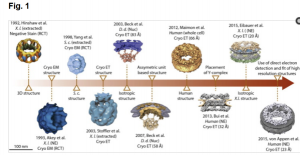
I rarely like to give praise to single papers because I know that the contributions of some individuals rests on hundreds of thousands of tiny “scientific breakthroughs” made by hundreds and thousands of individual researchers. But this paper has such detail about the nuclear pore and the protein positions that I just had to link to their article.  They graciously quoted several articles using cryo-em that have lead to further understanding of the structure-function relationship..

I am so eager to use this type of diagram and see what changes can be measured in the nuclear pore (from the myriad TEMs I have ) relating to such things as elevated “oxidative stress” and “in vitro” conditions.

Nuclear pore gallery-2

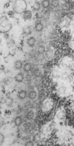
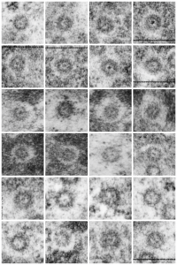
Adding more profiles of nuclear pores, from several different knock-out mice, basically to see whether the parameters I have set (chromatin exclusion zone, nuclear pore to nuclear pore distance, and distance between 20 nm DNA particles) changes with metabolic, or oxidative, or cell cycle (including apoptosis) stressors, and to see whether densities (probably pre-mRNA and other larger proteins that show up as occupants of the center of the nuclear pore) can be found to vary with similar cell states.  I subjectively choose the nicer looking pores, tending to bet mid-cuts, and some subconscious bias likely causes me to choose those pores with transport molecules, as here about 87% of pores have center densities, while when counted in a single micrograph (not my selection) the ratio of pores with and without central densities is closer to 50%.  we will see. The octagonal symmetry is very clear.

nuclear pores top down transmission electron micrographs


Measurements of areas surrounding the nuclear pore: hepatocyte


Some measurements (see previous post for comparison data), might be useful in a relative sense to others looking at nucleus, nuclear architecture, and nucleolar architecture.

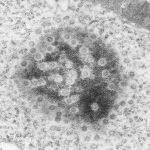
This particular image is unretouched tangential section of an hepatocyte nucleus (17529neg73229block, anm203,wcii -Gclc 28d).  There are numerous nuclear pores sectioned tangentially in this image, about 35 of which are there 15 or so in sufficient detail to demonstrate a density within the center of the nuclear pore demarcating the site that pre-mRNA and larger particles are ushered through (both ways) (about 44%). Compare this micrograph with a previous post of a similar tangential section for parallel measurement.  They are not identical, but close. Curley red lines demarcate polysomes.

In this micrograph the pore to pore distance is n=34 x=482+177nm, the distance from the outer part of the nuclear pore to adjacent chromatin is n=97, 62.7nm +/-32nm, and the distance between the 19-20 nm beads on a string in the chromatin next to the chromatin exclusion area is n=39, 46nm +/- 13nm. The actual measurements are shown at right (green=pore center to pore center), grey=chromatin exclusion areas, that is the distance between the edge of the nuclear pore and the adjacent chromatin, grey, and purple, the distance between the 20nm beaded-periodicity found in the pore-adjacent chromatin.

electron micrograph hepatocyte measurements of nuclear pore and exclusion area and chromatinelectron micrograph hepatocyte measurements of nuclear pore and exclusion area and chromatin