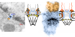
It is sometimes difficult to reconcile all the graphics that appear in research articles with what actually one sees with the electron microscope. There are artists and scientists trying to communicate facts and abstract ideas and the results are something hysterical.
Anyway, in looking at the 14CoS ko 48 hr no rescue nucleus in an hepatocyte I found this nuclear pore which actually had what looks like some of the 8-mer basket proteins that were quite clear. I have highlighted those (on the nuclear side of the pore) in blue and highlighted the cytoplasmic side with cytoplasmic filaments in rust color. One can see from a diagram from wikipedia (thank you wikipedia) which I have extensively simplified and modified, that the original and what I have seen in actuality don’t really fit. I cut the diagram apart and extended the inner nuclear membrane space, shrunk up the basket and extended the cytoplasmic filaments (see diagram on the right). The other issue is likely that the micrograph tends to show the pore from the ” cytoplasmic viewpoint” or bottoms up, while the diagram looks more “at the horizontal level”. Basically, the nuclear pore complex is an absolute wizard structure… fun to learn about. This electron micrograph is from a 48 hr old 14CoS ko pup, not rescued with NTBC. 18750_80206_anim#35 liver. More posts on this topic HERE.
 What i have trouble visualizing with electron microscopy are the three rings…. the cytoplasmic ring, the nuclear side ring and that ring structure that individuals claim is at the rounded fusion of the trilaminar inner and outer nuclear membranes.
Here is another nuclear pore from the same electron micrograph in which I have highlighted the four areas which perhaps will correspond to the nuclear side ring 2 dots at the bottom (along with light blue arrow of some particle just on the way out of the nucleus likely) and the cytoplasmic side ring above). Orange arrow points to where the trans membrane ring should be seen.

