Daily Archives: May 29, 2017

Explosive eruption: haha

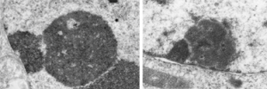
inner nuclear membrane nuclear protein traffickingHere are portions of two nucleoli, which just upon casual observation look like they are throwing up some granular components onto the inner nuclear membrane (particularly the image on the right). haha.  There are many  instances where a physical force appears to be exerted on the constituents of the cell (in this case the nucleus), I can mention two off hand, that is the bending-stretching-pulling look of mitochondria as they swerve over to become attached in some unknown respect to the intermediate filaments of the desmosome, and similarly  swerve to the outer nuclear pore filaments to connect there as well.  These motions in space have to have significance (at least in my own mind it would be silly to ignore them). At this moment in time I cannot tell you what this means, but one at least seems to be centered over a nuclear pore, the other, maybe a pore would be found out of the section.
 

Nucleolar ultrastructure: fibrillar center organization

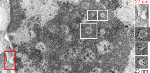
I was looking at this micrograph of a n hepatocyte with the fibrillar centers and dense thick fibrils in their midst, and the granular zone around the outside of this nucleolus. The latter being very punctate in appearance, just really obvious. I measured these once again at about 23 nm in diameter and noticed that around many of the fibrilar centers where was a basket, or ring, or radial distribution of these small punctate appearances.  OK so they are not really punctate, but can appear smeared like fibrils too, though the granular appearance and the nomenclature is well entrenched in the literature, they are just going to be strands not dots.  It could be that these radial dots around the fibrillar centers are bands, or windings at a given distance apart, kind of like fingers of a basket, or more aptly put, like a cage, with wires of 23 nm in diameter.

Electron micrograph here shows a portion of a nucleolus with three fibrillar centers cut out and enlarged (original sites are boxed in white), and a portion of the outer nuclear membrane with ribosomes (for measurement – one ribosome being about 27 nm in diameter, top right image with red dot and text). The cross section of the cage, or basket filaments around the fibrillar centers are shown as black dots.

Great humor: great truth

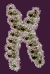
Reading about nuclear organization i ran onto this quote, and felt it was so great i just had to repost it. I personally continue to marvel at the naming of cellular structures, processes, and stuff, and the term “junk DNA” always rubbed me the wrong way, as does “non-coding DNA”.  I marveled to myself when reading those terms “what gives individuals the ego to believe they know enough to call anything “junk DNA”. I am showing a diagram of a chromosome (vector file with thousands of loops, and highlighted edges, iridescent graphic from a failed cover submission trial, a decade or more ago).

Well, here is someone who actually got that into a reputable journal… good for him — quote—from Thoru Pederson. Half a Century of “The Nuclear Matrix”, Molecular Biology of the Cell Vol. 11, 799 – 805, March 2000: “These kinds of ideas have been generally ignored because the noncoding DNA is so “uninteresting” as sequence (as if we were at present clever enough to be able to detect all “interesting” DNA text, which we certainly are not). At our present state of knowledge (ignorance) we can only view the noncoding DNA’s information content on the basis of what is absent [e.g., promoters, cap sites, splice sites, terminators, and poly(A) sites].”