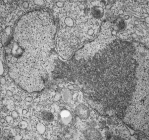
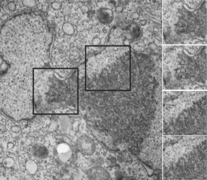
Here is kind of an interesting set of features in this clump of granular component of the nucleolus (or may just be condensed chromatin) that has a checkered appearance in some places (see checker boxes in inset micrographs to the right of the original image one showing the unretouched sections and an identical one with a checker pattern highlighted). There is a definite order to the spacing… or banding or filament pattern, something around 125 nm for the top two inset images, and for the bottom inset images, around 50 nm (which goes along with most measurements — being too big for interchromatin clusters granules (20-25nm), splicesosomes, around 30 nm, possible speckles? which are more like 50-100 nm).
Daily Archives: June 12, 2017
Nuclear pores: HeLa cells after irradiation with 30 joules UV
Nuclear pores: HeLa cells after irradiation with 30 joules UV — from an experiment trying to figure out how inhibitors of some of the caspases affected apoptosis. This study was from the 1980s and there are some interesting things never mentioned in that study which can be useful to someone out there doing electron microscopy on the nucleus.
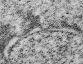
There can be no doubt that some controls on nuclear size shape and activities are changed in HeLa cells… probably so different than the HeLa cells which started out way earlier than that. But while looking over “nuclear pores” in a different study I just happened to look over nuclear pores in this HeLa cell which had been exposed to 30j of UV. I can find some evidence of nuclear pore cytoplasmic filaments but no nuclear pore baskets. So this is an N of 1, therefore not significant in and of itself, but when I googled nuclear pores and HeLa cells, there were publications, thus it is not an unusual, nor never-seen, entity.
Some of the central densities are missing from these nuclear pores as well.
 Other interesting features include the “presumed” granular portion thee is a pattern, both on the upper margin before the “neck” (whose significance is not known) and also on the surface past the “neck”, I have put a checker board behind elements which appear to be organized into alternating space. There are cytoplasmic filaments seen below but no nuclear side basket. This is the most perpendicular section seen in this micrograph.
