Monthly Archives: May 2017

Working out dimensions of nuclear and nucleolar components from archived electron micrographs of hepatocytes

It is nice to read that something is measurable, that has a dimension in nm that one can put into some kind of life-perspective. I don’t know how accurate those individuals were in giving a distance measurement to their images are, and I am giving each the benefit of the doubt in terms of attempting to be accurate) i guess they are quite accurate. I know for myself, using three or four different electron microscopes, and various different paper products, films, dryers, etc, that there is shrinkage, stretching, and all sorts of changes that have occurred over time (even 35 years) that affected the dimensions of objects on the micrographs, even though by a very small amount.

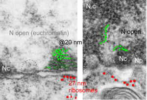
I am using 27nm as an internal control for magnification+enlargement of images. In this case, the 27nm red dot, in all images has been derived from about a dozen ribosomes, and labeled as such. The green dots are about 18-20nm in diameter in these micrographs, and strung as if “beads on a string” (not the beads on a string of nucleosomal DNA, but production of preRNA in the nucleus?) but is it not strange that there is a repeated motif – that is- beads on a string- showing up a lot, both in the nucleoplasm, the nucleolus and the cytoplasm (ribosomes on mRNA) as a kind of “convenient” little pattern.  And who knows where else this pattern might be found). So the green dots show this motif, in a parallel-linear pattern, in the condensed (or closed compartment) chromatin. Both of these examples come from Gclc ko mouse hepatocytes.  Red dots are ribosomes.
Chromosome territories and models of folded DNA

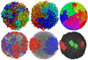
DNA folding: fractal globule and equilibrium globule renditions.  What awesome research and a daunting amount of data to handle. I read (or tried to read) one of the original papers by Job Decker  and another by Erez Liberman-Aiden but could hardly wade through them.  I am linking a blog called Explorable which was easier to read and recaps some of the figures in the original publications.

Altogether awesome, but I struggle that some the images which could actually use real electron microscopic (TEM anyway) structures so show these types of data…they don’t do so yet and there is much that could be revealed.  That would be a good way of bridging what is seen with the theoretical. The radial symmetry (on 2D bilateral using TEM) was obvious to me decades ago, not in the interphase cell so much as in the cell undergoing apoptosis… I am so glad this will be the case).

Here are a couple of images which show different models (fractal and globular) for DNA folding, and one that shows the radial symmetry found in the more_transcribed (which i would dare to call euchromatin, and lesser_transcribed (which again, i would dare to call condensed chromatin) regions of the DNA. I had always kind of thought that the active regions would be at the interface between those two TEM visualized domains…particularly at the nucleolus.

What haunts me still is what I saw 50 years ago in tissue culture is the spinning nucleus (time lapse photography), I have yet to have anyone put that into the puzzle, for a motion and distribution to the nuclear pores, of newly transcribed RNA.  Certainly that is in the mix somewhere, unlike these static models.  Below is a shameless cut and paste of the several images that come up on google for DNA chromatin territories (some fractal globule, some equilibrium globule, far right lower, green is more highly transcribed region of a chromosome.)… AND where is the nucleolus.


Explosive eruption: haha

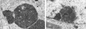
inner nuclear membrane nuclear protein traffickingHere are portions of two nucleoli, which just upon casual observation look like they are throwing up some granular components onto the inner nuclear membrane (particularly the image on the right). haha.  There are many  instances where a physical force appears to be exerted on the constituents of the cell (in this case the nucleus), I can mention two off hand, that is the bending-stretching-pulling look of mitochondria as they swerve over to become attached in some unknown respect to the intermediate filaments of the desmosome, and similarly  swerve to the outer nuclear pore filaments to connect there as well.  These motions in space have to have significance (at least in my own mind it would be silly to ignore them). At this moment in time I cannot tell you what this means, but one at least seems to be centered over a nuclear pore, the other, maybe a pore would be found out of the section.


Nucleolar ultrastructure: fibrillar center organization

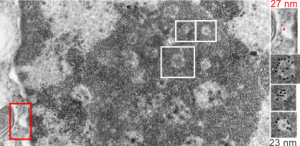
I was looking at this micrograph of a n hepatocyte with the fibrillar centers and dense thick fibrils in their midst, and the granular zone around the outside of this nucleolus. The latter being very punctate in appearance, just really obvious. I measured these once again at about 23 nm in diameter and noticed that around many of the fibrilar centers where was a basket, or ring, or radial distribution of these small punctate appearances.  OK so they are not really punctate, but can appear smeared like fibrils too, though the granular appearance and the nomenclature is well entrenched in the literature, they are just going to be strands not dots.  It could be that these radial dots around the fibrillar centers are bands, or windings at a given distance apart, kind of like fingers of a basket, or more aptly put, like a cage, with wires of 23 nm in diameter.

Electron micrograph here shows a portion of a nucleolus with three fibrillar centers cut out and enlarged (original sites are boxed in white), and a portion of the outer nuclear membrane with ribosomes (for measurement – one ribosome being about 27 nm in diameter, top right image with red dot and text). The cross section of the cage, or basket filaments around the fibrillar centers are shown as black dots.

Great humor: great truth

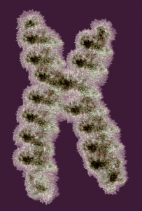
Reading about nuclear organization i ran onto this quote, and felt it was so great i just had to repost it. I personally continue to marvel at the naming of cellular structures, processes, and stuff, and the term “junk DNA” always rubbed me the wrong way, as does “non-coding DNA”.  I marveled to myself when reading those terms “what gives individuals the ego to believe they know enough to call anything “junk DNA”. I am showing a diagram of a chromosome (vector file with thousands of loops, and highlighted edges, iridescent graphic from a failed cover submission trial, a decade or more ago).

Well, here is someone who actually got that into a reputable journal… good for him — quote—from Thoru Pederson. Half a Century of “The Nuclear Matrix”, Molecular Biology of the Cell Vol. 11, 799 – 805, March 2000: “These kinds of ideas have been generally ignored because the noncoding DNA is so “uninteresting” as sequence (as if we were at present clever enough to be able to detect all “interesting” DNA text, which we certainly are not). At our present state of knowledge (ignorance) we can only view the noncoding DNA’s information content on the basis of what is absent [e.g., promoters, cap sites, splice sites, terminators, and poly(A) sites].”

False advertising? Kroger consistently does this!

Why does it make me so mad when i see a new advertisement on a Kroger product that I know has had the price raised? I have heard various explanations from Kroger personnel when I ask them.

Case in point: Here is a new price for Kroger chicken breasts, which were $1.99 per pound and the price was raised — but rather than just being “up front” they have to couch the new price in a fake news sign that says LOW PRICE, when in fact it is an increase of about 10%.  Do they think that all customers don’t notice. Do they think that i has no impact on their overall credibility.

Who does this kind of advertising? No one can do this and also run an honest business.  NO ONE.  THIS IS FRAUD, on the level of Kroger, many current businesses, and also the running of Washington.  Big business, and big government run like a business, has lost all sense of morality, and have license to lie, coverup, and falsify..perpetrating bad food and bad governance on the US.

Kroger company needs to have its CEOs sent back to school in honesty and integrity, not profitability.

An honest approach would be so refreshing… something like… “to our loyal customers: we are sorry that we have to raise the price on this product, we hope you continue to buy it” 

Estimates of the size of some nucleolar components

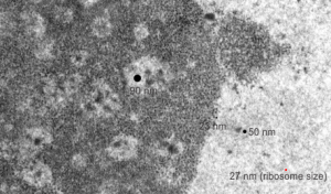
This micrograph, parts of which have been seen before, is an hepatocyte nucleus which is in the early stages of demise (untreated CoS14 ko without rescue). The nucleolus is large, and has two cajal type bodies (one seen here) and fibrillar centers with fairly large densities within (of approximately 90 nm diameter). I have given these sizes (relative to a ribosome size red dot approximately 27 nm (lower right hand side of micrograph labeled as such). The granules within the granular portion of the nucleolus (as well as the banding patterns found on cajal bodies) is something around 23 nm.  What I presume to be perichromatin granules look to be about 50 nm. The large dense bodies within the fibrilar centers are a little unusual.

Interchromatin granule cluster ?

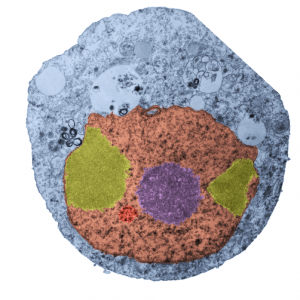
Nothing is black and white, except electron micrographs, and even then they are easy game for dodging, blending, blurring, mending, and pseudocoloring. Cytoplasm here is blue. In this case I have a cell from a culture of A549 cells in which C9orf82 was knocked down using si67.  This is pellet 4 from one of those studies. It produced this cell in which the granular component of the nucleolus was separated into at least two bodies (almost bilaterally symmetrically arranged) which rest at the inner nuclear membrane. Fibrillar centers are small, dense fibrillar component is still visible (albeit not distinctly) in the nucleolus (pseudocolored purple in the center of the cell).  Off to the side is what I could guess would be either degraded DNA (not my first choice as an answer (it is pseudocolored bile-yellow) or perhaps a fine granular component of the nucleolus in an apoptotic cell.  Part of an interchromatin granule (red, lower left) cluster (IGC) (sometimes called speckles, a name which is not really to my liking since the IGC has a boundary, a background texture that is also part of the structure).  So I would propose keeping two names, the “IGC” which is the greater boundary of that area and “nuclear speckles” as well, since the densities (as seen in this micrograph) can apparently be large.

If i am not off base here, the background of the whole IGC is red, but there are very clearly larger than typical speckles within the IGC, and the latter itself is quite small.  With immunohistochemistry the diffuse staining of IGC background proteins fluoresces with some of the antibodies to proteins like SC-37 in an area which can be 2 microns across easily.  I am looking for alternative proteins stained in very punctate regions of the IGC and hopefully they will be different and good markers for a separate ultrastructural components of the IGC. The point would be to localize to the densities with the IGC, some proteins for splicing, and accept that the diffuse staining could apply to SC-37, but not other proteins, mainly those in the variable size granules within the IGC.

We will see if there are data to back up the keeping of both names but assigning them to their obvious separate entities. BTW, there is pretty obvious bilateral symmetry to this apoptotic cell nucleus. and a radial symmetry to the dense bodies within the red colored IGC. And while i have no clue what the curley-Qs are… i bet they have something to do with the transfection. Just looking at this cell shows one tiny surviving mitochondrion, in this apoptotic cell, which is very close to the cytoplasmic filaments of a nuclear pore…also something which needs to be monitored during apoptosis.