Daily Archives: June 27, 2017

More nuclear pore measurements

This hepatocyte was from a mouse (#5) which was a 14CoS nujll that did not receive NTBC as a rescue drug. This is 24 hours into the neonatal period.  Nuclear pores have fewer central densities (presumably being proteins transported). I am measuring for distance beside the nuclear pore that the chromatin exclusion zone hoping that differences will show up with various conditions in previous experimental models.

Top micrograph, unretouched, bottom micrograph, pores used in calculation have circles, red dot = a 27nm ribosome, purple dots are areas of the chromatin just adjacent to the chromatin exclusion zone surrounding the nuclear pore.

meaurements of the chromatin exclusion zone around the nuclear pore transmission electron microscopy

Nuclear pores and polysomes and other things

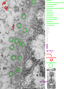
Continuing an evaluation of nuclear pores in various experiments from the past, here is a microgaph from 17709_65053_14CoS-/- NTBC 1yr. I used some of the opportunely sectioned “almost top down” cuts of nuclear pores to look at the inter-pore distance (which here measures , the chromatin exclusion zone, and the distance between the adjacent chromatin areas that have that “beads on a string” look.  Distances are derived from an approximate size of the ribosome at 27nm in diameter, nuclear pore diameter of 120nm, and a 19nm diameter for the adjacent chromatin at the edge of the chromatin exclusion zone.

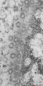
Unretouched photograph of portion of a nucleus from an hepatocyte from CoS-/- mouse maintained on NTBC for 1 year.  and the marked-up micrograph below.

The nuclear exclusion zone is 54nm+18, the inter-pore distance is 289+119nm, and the distance between the densities in the chromatin (purple dots) of the edge of the chromatin exclusion zone is 46+5. Red curly-cues are polysomes, red dot is one ribosome (taken at 27 nm). Green 8-edged rings are nuclear pores. (bottom picture)