Monthly Archives: July 2017

Liquid breathing E2: 3 hr 17 days recovery

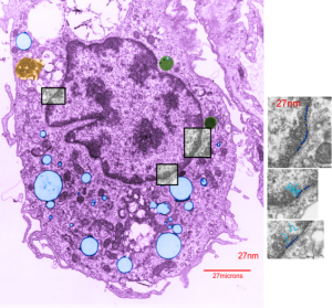
Here is an electron micrograph of an alveolar macrophage from a mouse that liquid-breathed E2 for 3 hours and allowed to recover for 17 days. The droplets of E2 (which i pseudocolored blue) are well defined and surrounded by a relatively prominent membrane and likely have some characteristics of lysosomes or endosomes because of the peripheral electron density. Two multivesicular bodies/late endosome type structures (pseudocolored green) show tiny bits of E2 (blue) as well (at least this is how i interpret it). This was sort of typical for E2 droplets in similarly treated mice.  Organelles within this alveolar macrophage seem relatively healthy  and importantly, the condensed chromatin is typical of non-activated macrophages in general. There is some endocytic function remaining as there is an inclusion of tubular myelin (orange) contrasted with the pink nucleus and cytoplasm.

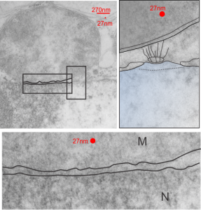
The three grey insets (notice that the bottom one has been mirrored horizontally) show a distinct periodicity to the most peripheral chromatin against the inner nuclear membrane.  And there are places where the size and spatial arrangement of the densities change (smaller “somes” – whatever state this condensed chromatin is in with its attendant RNA and other proteins).  The spacing (a little smaller than the dark blue dots) is something close to 30nm when I use a mean diameter of a cytoplasmic ribosome as the measure of 27nm for this image. 1432_mouse_lung_liquid_breathing_3hr_17d_recovery.
Little green aliens among us

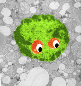
So sorry, ha ha, but when i see this kind of nuclear symmetry (an electron micrograph, with two little presumptive eyeballs staring at me (condensed chromatin), zygomatic arches coming off (as condensed chromatin on the sides of the nucleus), and a hairline part at the center of the frontal bone (condensed chromatin and nuclear pores on the inner nuclear membrane at the top of the nucleus) and a pointy chin, I am compelled to laugh out loud and add this to my collection of TEM-Devils, and sometimes other objects like hearts and ghosts and snowflakes. Anyway, here is a little green cutie, which spawns from a mouse hepatocyte null for 14CoS and not receiving NTBC as a “saving” medicine and prone therefore to undergo massive hepatocytic apoptosis within a day or so of birth, and ultimate demise. This mouse is 24 hours post partum and showing signs of hepatocyte apoptosis, and mitochondrial and ER disarray, neg 16042, block 65718, anm#5, one chatter line removed with photoshop, layers constructed in photoshop, pseudocolor and eyes added in CorelDRAW.  Dont you wish there was a place to publish such stuff…. Maybe i will start my own journal…  help me think of a name.  (LOL)


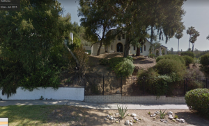
5252 Windermere Ave, Los Angeles California 90041

This is where i grew up, so sad, the cute three-arched window in the master bedroom (which was small) has been stuccoed up, and the yard looks like no one cares.  My mom and dad took great care of this place which they purchased for 17k from my dad’s mom probably sometime in the late 1940s.  My brother and I did the yard on saturdays before going down to the Cinema on Eagle Rock Blvd. for a 10c movie.  I earned the money for the movie by raking and digging up dandelions from the grass. There was a loquat tree in the back yard which we climbed and ate from. A crawl space where my brother and I had a Lionel train set up with tracks and miniature houses, and props that we built, and a work bench with a really nice bench jig saw (the kind with an arm) and tools and a place to build model airplanes.

I walked back and forth to school often, and also from college  (Occidental College was only about a mile+ away, my dad dropped me off there on his way to work early in the morning). I lived there At least 23 years there, then the family moved to Hill Drive, just around the corner.

It was a tight squeeze for 5 children and two adults in this small house, (3 girls in a bedroom that need specially made small beds to fit against the wall, two brothers in bunk beds in the adjacent room) and there were sometimes cats, and a dog or two, and a neighbor’s child who was young when her mother died, and my mom kept her after school.

But it was big enough for Cub Scout meetings, and  Blue Bird meetings, and make believe dramas in the back yard as Campfire girls. A hand made play house, with a roof and windows –big enough to walk in, and some boards in the neighbor’s tree for a platform, a swingset, from which a neighbor boy jumped, breaking his arm, a place for other mischief and ouch, when my littlest sister wound her pinkie up in a bicycle gear.

Below the house on 5252 Windermere is the house which my music and art teacher lived, Mrs. Constance Braasch.  She had a tremendous influence on my life.  I still (after 70 years, play piano, she was my teacher for at least 12 years) and have a couple pictures and china pieces that we painted together.  She was a remarkable woman.

Outer nuclear pore filaments and mitochondrial proximity

These little junctions, that is, nuclear pore – mitochondrial associations, have been fascinating to me.  I have noticed and photographed (whether ideally fixed, or in focus, or in this species or that cell type, just as many as i can) to try someday to figure out what tasks they are carrying out. The obvious of course, providing energy, maybe for nucleo-cytoplasmic transport, but maybe too for nuclear rotation, as we all know happens from observing those old time lapse videos of tissue culture cells with mitochondria dancing close to the nucleus and the nucleus spinning  — ha ha – new question, to nuclei on the northern hemisphere rotate the same way as nuclei cultured in the southern hemisphere?) and also for nuclear shaping, but importantly, do they have something to do with chromosome territories? That is, something more than just an indirect influence like shape or transporting molecules.  Might they participate in the transport of ions through the outer pore areas as well as the core.  Might they influence assembly and repositioning of nuclear pores, making and or breaking them.

This particular nuclear pore-mitochondrial association is not the best micrograph in the world by a long shot, but it is from a heterozygous animal conditional ko of  Gclc in the liver (wc/ii) so has some increase in oxidative stress.

The original micrograph is on the left taken at 9500x, neg 17535 block 73458 anm# 505 wt hepatocyte.  filaments (wiggly lines) going up toward the mitochondria (top shaded grey) are prominent , the nuclear basket is not prominent, and something looks to be a pattern underneath the pore (black oval) that would likely be some tangential portion of the nuclear membrane (not hazarding a guess as to whether outer or inner.


Ciliary glycocalyx or crown

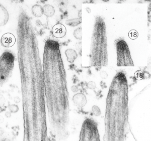
I just found these nice electron micrographs digging through the stack of 20k images and they are of the stomach of a mouse that is null for the gastric HKAtpase alpha. This was part of a study long time ago with many other main authors. The tips here are in the stomach, which was one of those unusual findings, that is, ciliated cells in and among the parietal and zymogen and mucus cells.  Clearly ciliated however.  So the tips of these cilia are really nicely preserved and in focus and it is clear that there is a glycocalyx, not exactly like that of the brush border in the intestine, more like that found in the trachea, typically “normal” ciliated epithelium. So below are TWO images (one inset) tips of 5 cilia which clearly show 6-17 filaments, and my projected total is given as well.  The electron dense  “sphere” or “dome” of attachment subadjacent to the plasmalemma is very obvious. 16447 and 16448 block 65165 gHKAa-/- mouse

I googled images to see what current research had been done on these fine filaments and the density seen within the tip of the cilia in the first figure below and found a reference back to 1978 (Charles KuhnIII, Wayne Engleman The structure of the tips of mammalian respiratory cilia Cell and Tissue Research January 1978, Volume 186, Issue 3, pp 491–498) which put the estimate of the number of filaments at something around 6 or 7, which makes no sense to me in 3dimensions…. So in the circles I approximated what i think the number of filaments would be. I found a second “antique” paper (only calling them antique because I have papers that old as well which contain details not really found about ultrastructure in current literature) and it also thought the number of filaments was in the single digits. This diagram by Spicer (which of course I would redraw with more filaments). It is cute.

Sadly, a more current paper also quotes a low number of filaments… but has an interesting observation about a periodicity, so a quote from them is HERE “The apical structure of human respiratory cilia was studied using thin and thick sectioning techniques. The distal end of the mature cilium is narrowed and presents dense material and a crown-shaped glycocalyx. The crown is composed of three to seven bristles, having a mean length of 31 nm and showing a periodic structure. The capping structure of the apical part of human respiratory cilia is composed of a plate and an electron-dense cap tightly bound to the central and to each of the nine peripheral A microtubules. Lateral spokes link the peripheral microtubules to the internal ciliary plasma membrane and dense filaments radiate from the dense cap to the ciliary crown. These ciliary apical structures may be involved in several functions associated with microtubule assembly as well as ciliary movement and mucociliary transport. Apical structure of human respiratory cilia. Available from: https://www.researchgate.net/publication/19463484_Apical_structure_of_human_respiratory_cilia”

@70 degree separation of homologous chromosome densities

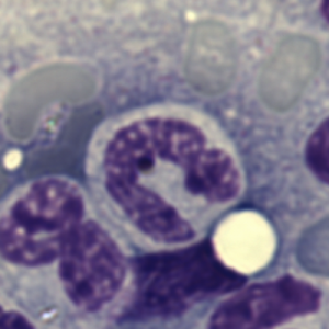
The nuclear center might not be a simple concentric center.  Why, maybe to allow for stretching and shape changing, particularly in leukocytes and migratory cells.  Seems like a pretty smart approach to something that ‘can be round’ when it is physically convenient, but is more likely to be strung out pole to pole along with the cytoplasm, or independently of the cytoplasm as well (again, leukocytes, particularly the polymorphynuclear leukocytes).  This image of a bone marrow cell from mouse has an asymmetrical shape, which is easily found, in practically any microscopic field or any bone marrow smear. Maybe like a U, but more a V really as the chromatin densities are asymmetrical in this view and in many views. Even if one takes into consideration that this is still 3D (even after spreading) and that there might be two condensed chromatin areas where there appears to be just one in this picture, the U as a whole shows a bilateral symmetry maybe in two dimensions, but not in the third dimension. This begs the question what are the symmetries in x,y and z that unfold as leukocyte nuclei become lobular.

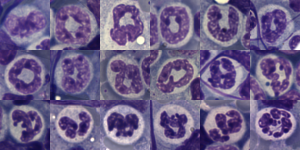
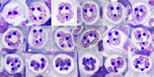
The following two images (composite markups from bone marrow smears from a single mouse. I should look up the treatment protocol, but i am pretty sure because of the number of images that it was receiving BaP.  Not all nuclei are identified as belonging to polyymorphonuclear leukocytes, but what is common is two or four sets of condensations (obviously not all can be called the Barr body chromosome drumstick) but the pattern is clear 1] bilateral left to right x axis, bilateral in the Y axis (narrow bottom two definite condensation sites at the top, and a C shape (backwards) in the z axis shown in a group of progressing nuclear lobulation, and below that, lightened images lying under dots over areas of chromatin condensation … I tried to orient the wider separation of the sister condensation points toward the top.


Areas of the nucleus where chromatin organization will differ.

Areas of the nucleus where chromatin organization will differ:
These I can think of that could have different morphologies; just in the Gzero and  the resting phase of the cell cycle.  Presumably all these areas it is DNA plus their manditory core proteins (histones etc splicing complexes polymerases and so on).

condensed chromatin adjacent to the nuclear lamina (two phases)
condensed chromatin at the hemisphere around nuclear pore complex
condensed chromatin from the Barr body with complete silencing
condensed chromatin from some epigenetic silencing
condensed chromatin at areas of nearly complete silencing
condensed chromatin and RNP of the granular component in the nucleolus
condensed chromatin and RNP of the dense fibrillar component
euchromatin within the fibrillar center (nucleolus)
euchromatin within the interchromatin granule clusters
perichromatin granules (and their adjacent lucent ring areas)
condensed chromatin and RNP in nuclear speckles
degree of cell differentiation and areas of ‘perhaps’ digestion

An additional array of changes would certainly occur during the mitotic cycle, and during the process of apoptosis while chromatin is being disassembled.

Arc of separation between homologous chromosomes? angular dimension?

Arc of separation (degrees of separation) , or central angle? which ever one should call it, i don’t know, between chromosome pairs in their respective chromosome territories. I see it everywhere in the FISH and other fluorescent imaging of chromosome territories. I see it in bone marrow spreads as well, clearly shown by areas of chromatin condensation of certain chromosome – or portions thereof.

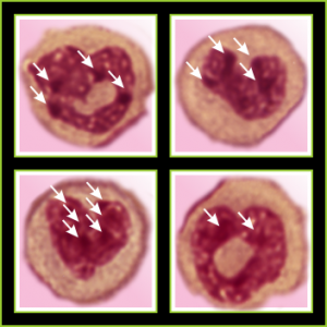
So here is a bone marrow cell which has obvious chromosomal densities, and I am presuming that these are condensed by epigenetic or other influences but there is definite symmetry (in 3 axes) and differing angular dimensions to the presumptive pairs of chromosomes. The whole nucleus is in a V shape, the point of the V being “down” here but in terms of 3D space, it may not be. It certainly represents a portion of the nucleus around which the symmetry occurs. Simetimes instead of being seen as a “v” (or heart as I have depicted it here) a U is present. and this might represent a less well condensed form of the same type of compaction, on the way to becoming a multilobular white cell.

The arc drawn between what is felt to be homologous areas of condensed chromatin, billateral symmetrical dark spots (highlighted in black on images to the right of the unretouched (but color adjusted to red) giemsa stained white cell).  The arc is influenced of course by random position of the cell as it is smooshed onto the slide during preparation as well as the spot identified as the center of the nucleus… whatever that might turn out to be, but i bet it is somewhere near the apex (bottom of the heart).  The actual physical center of the nucleus is not well defined in the literature.  So i think the concept of radial chromosome territory positions might need to be altered to account for the z axis of symmetry and the position which ultimately will be described as the nucleolar “center”… which cant be the nucleolus only, since often there are multiple nucleoli…  maybe the term center of the nucleus should be called “centers of the nucleus” and the arc of separation determined from various sites.

Love in any language

Totally unnecessary post of bone marrow nuclei: but what it says very clearly is that symmetry prevails, may not be the symmetry we expect from a set of identical chromosomes all lined up in on-off status, both sides the same (X chromosome or barr body excepted).  I see here a great symmetry, right to left mirror, top to bottom asymmetry, pointy at the bottom because this is an area where the nuclear membrane might just get segmented into a thin band between two bottom areas of chromatin…. going from what might appear to be a point, to two cheeks.

I am thinking about writing up an editorial on this symmetry…. make the cover submission first, always my motto.  Each of the hearts below is from bone marrow from an hypoplastic animal (CyP1a1null) either given benzo(a)pyrene or control diet.  These particular nuclei are not yet hypersegmented…  just fun examples of symmetry, which i have outlined in an identical micrograph below this.

four bone marrow nuclei polys chromatin condensation symmetryfour bone marrow nuclei polys chromatin condensationSo there is symmetry, it is bilateral in one dimension, but will take some thinking to see how to describe a third dimension in symmetry.

Because the chromosome territories likely (per published data) kind of interdigitate, most assuredly, at many sites along each chromosome for functional reasons) the symmetry will be highly simplified in light micrographs at best.

All this discussion could lead to a place to examine the chromatin patterns in the X (barr body) in neutrophils where they are hanging on by a nuclear membrane thread – so to speak) and other parts of the neutrophil nucleus where transcriptional activites are going on.

I found a match for what I see in bone marrow… yippie

For at least one decade I have thought about some nuclear morphology, typically found in bone marrow in progenitor cells (mouse) particularly in mice who have been dosed with 125mg/kg/day benzo(a)pyrene (BaP) for 18 days, underpinned by the presence or lack of Cyp1a1 (Fig. 3. Histology of bone marrow) which showed severe hypolasia of the bone marrow and a significant increase in the hypersegmentation of the bone marrow cells (all kinds). Lymphocytes were increased regardless of BaP in the Cyp1a1(-/-) mice regardless of BaP treatment as was hypersegmentation. So this was interesting, but never pursued as a separate topic. I have micrographs which show this same bilateral segmentation, which may turn out to be portions of (or whole) chromosome territories. I searched the literature (the relatively current literature and current literature during the study and didn’t find anyone who had any images which would suggest the cause.

Then as chromosome territories became a target of interest, two very old (I mean really old) documents were cited in Cold spring Harbor Perspectives in Biology in 2010: (Cold Spring Harb Perspect Biol. 2010 Mar;2(3):a003889. doi: 10.1101/cshperspect.a003889. Chromosome territories. Cremer T, Cremer M.) listed as Rabl 1885, and Boveri 1909 (In the drawings of two cells in prophase from Ascaris megalocephala univalens embryos. He mentioned that these protrusions were germ line chromosomes.  This renewed my interest in trying to figure out which chromosomes were blipped out (or cued for hypersegmentation) in the null mice, and whether this indirectly relates simply to high levels of cell renewal in their hypodcellular bone marrow or whether it relates directly to the loss of CyP1a1 in mice somehow.

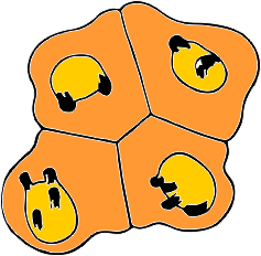
I have redrawn the figure of Boveri (1909) and added some images from bone marrow of hypersegmented cells (which I now assume to be specific chromosome territories of specific chromosomes, but which chromosomes is not known. Another interesting point is that Boveri thought that the two top cells were initially responsible for the two bottom cells (which he called daughter cells) and the difference in two vs four areas of chromosome blipping are evident (this would be high condensation and sort of a promise later of becoming an area tethered by a thin strand of nuclear membrane, or budded off as a microbody).  Certainly this man spent a lot of time at the microscope.  These types of symmetries, and associations are randomly seen usually and lots and lots of view-time are required before patterns emerge.

(Adding one more coincidence? comment! is that Boveri drew his cells with about a 64 degrees  of “arc” from approximate (obviously not know for sure –i dont even know if anyone has decided where the “center” of the nucleus actually is.. ha ha–i will have to google that). When i calculated the angular dimensions of the two similarly painted chromosomes in FISH and other fluorescent tags for chromosome territories, i could almost always envision and measure a distance of about 70 degrees between chromosomes.  This is kind of exciting, as it give bilateral symmetry to the nucleus which is NOT an exact mirror,  but something different.  The Arc of Separation between chromosomes I will have to research.