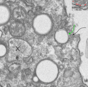
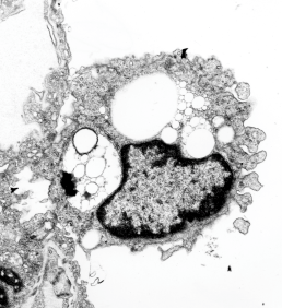
The bounding electron dense coat of proteins on the perfluorochemical droplets, whether phagocytosed from a neat liquid as inhaled from liquid breathing, or provided to the blood stream as artificial blood, it seems to be a fixture of the morphology. I have scanned the edge of a single alveolar macrophage for evidence for “point of entry” of a droplet of E2 into said cell. It is pretty clear even from this slightly too thick, thin section of a mouse lung (1349 4840 E2 3hr 48hr recovery) that the electron dense coat around droplets exists in lung of liquid breathing mice before they are phagocytosed by alveolar macrophages. In this case the proteins likely to be lipid molecules derived from surfactant.
I suppose it is also likely that the slight lipophilicity of perfluorocarbons means that the surfactant lipids (such as phosphatidylcholine and phosphatidylglycerol) and the amphipathic proteins in surfactant (like SP-B) might contribute in some way to this droplet coat (as derived from surfactant already in the alveolus) reflecting its contribution shown in vitro studies suggesting an ability uptake of bacteria from the alveolar space (the leap is to E2 droplets) (ref Yang et al below)
There are at least 4 whole E2 droplets, and two cut off (left hand side of micrograph) droplets of E2 in this image. The tiny dotted periodicity were shown by lines in the droplet at the middle right, the green arrow points to the electron dense bounding layer of “some lipid and protein components from the alveolar space, likely from surfactant”, and the black arrow points to the space in the plasma membrane where the particle is likely being engulfed by the alveolar macrophage. Ribosome size is judged by the upper right corner inset with a portion of RER enlarged identically with the larger micrograph. X marks an inclusion type that I have seen before but do not recognize. Any suggestions from the audience are welcome (millermn – ucmail dot uc dot edu).

Surfactant protein B and lipids look like a good candidates for the electron dense cover for E2 droplets — reading the article (Hawgood et al) and Yang et al specifically referring to SP-B and from which I quote: “In no rank order these activities include membrane binding, membrane lysis, membrane fusion, promotion of lipid adsorption to air–liquid surfaces, stabilization of monomolecular surface films, and respreading of films from collapse phases.”
SP-B has some interesting properties as referenced in many articles in PubMed which suggest that it has saposin like properties. Wikipedia identifies saposins thusly: “Saposins are small lysosomal proteins that serve as activators of various lysosomal lipid-degrading enzymes”. It is interesting that the huge lysosomal response that is induced by liquid breathing both in alveolar macrophages (and also when perfluorochemical emulsions when given IV) might be seen in the types of lysosomal bodies found, the length of time required to degrade/and/or reassociate the enzymes that appear to cover the droplets in the alveolar space, and concentrate (add more? or recycle more? possibly be reflected in the fine granularity of the electron micrographic views of these MVB/LE/PFC organelles. The amount of re-emulsivication of phagocytosed E2 in alveolar macrophages appears to be time dependent, and would fit with the tim required to increase in production of some lysosomal enzymes and become associated with the E2 droplets.

