Daily Archives: August 29, 2017

The right answer pushes the wrong button

endless monotonous skipping track
derelict emotion everybody loves me
enter the vein
dark electric absence
pilfer the flow of awareness
to buy your silence
you know the stillborn fantasies
memories of that which never happened

a realization fries the skin
crunch-perfection sizzles in the
grease of human conflict
manipulated and molded
ensuring fascist perfection
self propagated goodness
with a professional appeal

the right answer pushes the wrong button
immortal decay shows her chaotic smile
leaves only the inconsequential choice of path
the model number and price of inevitability
time is money to purchase a longer more elaborate death

AEMiller @1995

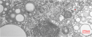
Variety: the whole spectrum of lysosomes/inclusions of E2 in an alveolar macrophage

Variety: the whole spectrum of lysosomes/inclusions of E2 in this micrograph of an alveolar macrophage (at least the whole variety I have seen so far). It includes lysosomes (LE/LY/PFC) which probably could be classified all the way from golgi vesicles to late endosomes since it seems that PFCs in general are able to work their way into the whole ER system (though to date I have not seen anything that looks like a PFC droplet in any ER which has ribosomes….  still looking). There are droplets in a “hollow” meaning no lysosomal enzymes yet, on the right but still having E2 droplets with the coating they must pick up from somewhere (maybe alveolar surfactant in the alveolar space?, being lipid and E2 being slightly lipophilic), to the dense small lysosomes with many E2 droplets on the left, to a single E2 droplet on the lower left, and a dozen droplets which look like they are hanging out in the cytosol with no membrane boundaries, to bottom center structure which looks like two E2 droplets in a lysosome with a lamellar type surfactant inclusion.

There is no paucity of free ribosomes in this cropped image, and size can be inferred from the approximate 27nm diameter of a ribosome (red dot), to the larger droplet size (bar=270nm).

Countless PFC droplets in alveolar macrophage lysosomal body

The lysosomes in this alveolar macrophage from a mouse which breathed E2 for 3 hours and then allowed to recover for 5 days are awesome.  The macrophage has produced enzymes which re-emulsify the E2 into very small (in most cases) droplets within the lysosomal structure. In addition, the enzymes make a border, which is very difficult to distinguish from the trilaminar membrane which surrounds the lysosome proper.  I don’t know how to explain this look of a double membrane, but figure it is partly a physical interaction between the E2 droplet (and not unique to E2 but seen with many other perfluorochemicals) and its hydrophobicity, and slight lipophilicity.

The smallest spheres of E2 are down around 35nm diameter, and size can be compared to something just larger than a ribosome (@27nm). Picture on the top is the unretouched (i may have removed a scratch with the band-aid tool in photoshop but nothing else), and it is not too great an image (scanned from the 31/4×4 acetate negative), but interesting still, and providing lots of information. Image below is one where I have highlighted the E2 droplets in a single membrane bound lysosome and embossed them.

The dark structure (also rounded) in the upper right hand corner of the images is what I think is a phagolysosome that contains mostly phagocytosed surfactant lipids (some layering and myelin-look is seen within this structure) from the alveolar space. There are also two mitochondria near center top. It also seems likely that some of the surfactant engulfed by alveolar macrophages would find its way (during re-purposing or re-cycling) into such lysosomes containing E2 droplets.

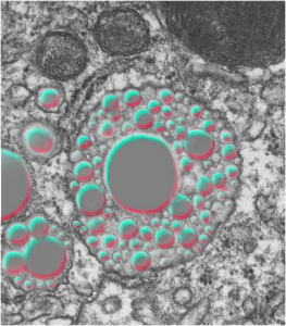
electron micrograph alveolar macrophage E2 liquid breathing perfluorochemicalelectron micrograph alveolar macrophage E2 liquid breathing perfluorochemical PFC highlighted