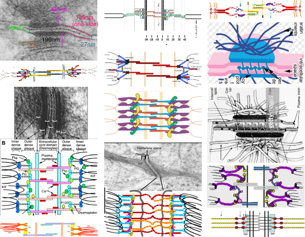
Desmosomes are kind of like Oscar rivets, and are blind perhaps, or double-sided. the latter have splits (the desmogleins and desmocollins) along the shafts. These splits cause act like flares and join in the central dense line (periodicitities) of the desmosome. The plakoglobins and plakophilins form the “head” of the rivet along with desmoplakin, and these rivets are unlike most rivets found for metal and wood work, because they are flexible, and have a “spring-hinge” at their head, the desmoplakin-intermediate filaments junction (which i believe is perpendicular not parallel (as most of the diagrams show), which can flex and bend and allow for motion of the desmosome as it gathers together as weld between two cells.
Below is a set of diagrams taken freely from the internet from publications on desmosomes (including my own). It can be seen that the majority of diagrams don’t really “see” what the microscopist sees as the course that intermediate filaments take when they are adjacent to desmoplakin. The diagrams on the lower left, and all those in the center and the lower right have intermediate filaments coming from the cytoplasm and making hairpin turns at the desmoplakin molecules…. However, the micrograph on the upper left, and the diagram in the upper right have the intermediate filaments which actually appear to arc ever so slightly, to be NOT forming hairpin turns to desmoplakin, but gently curving perpendicularly to the desmoplakins.
It seems more likely that the type of shear stress (the connection between desmoplakin and the intermediate filaments perpendicular to one another) would be more advantageous at binding two cells together than the diagrams below (with hairpin turns) which suggest that the connections between intermediate filaments and desmoplakin are parallel, thus involved in tension stress. The former intuitively suggests more efficient shock absorption over a smaller distance than the latter, and actually looks more like the actual anatomy than the latter.
The disparities among the images from electron microscopy and the diagrams from the literature are the reason that I began to try to think this through. Intermediate filaments are seen in micrographs that look perpendicular to desmoplakin (like what is seen in the micrograph in the upper left part of the diagram as brown lines), or as 10nm round dots (if they are cross sectioned). Any ‘apparent’ long intermediate filaments parallel to desmoplakin (as seen in the bottom center electron micrograph) can almost always be explained as tangential cuts.
This arrangement is most clearly demonstrated when intermediate filaments form a flat perpendicular band between the desmoplakin molecules of the desmosome and the mitochondrion when the desmosome and the mitochondrion are tethered together.