Daily Archives: January 5, 2018

Desmosomal-mitochondrial tether Stub tail monkey hepatocyte

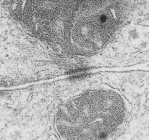
This particular electron micrograph of a desmosome with mitochondria tethered to either side shows some nice orientation and detail. Particularly the intercellular space has the zipper lines that are the desmocollins and desmogleins. These lines have some regularity, but owing to the enormous numbers of possible orientation that one could get in TEM, it is not that likely that a perfect one will ever arise. I even consider the roundness of the “spot” desmosme and the possibility that the organization is radial, wouldn’t that be fun. Someone out there with 3D imaging skills could certainly test this with the molecular models that do exist.  I think it would be just as fun as looking at the tomographs of thicker sections.  Brown dots are likely areas where desmocollins and desmogleins are intersecting-interacting, and these represent the intercellular central dense line of the desmosome. Black lines are areas where the 5-repeats in desmocollin and desmoglein (i suspect) are spanning the intercellular space. The black dots are some kind of periodicity visible on the outer lamina of the trilaminar plasmalemma.  I didn’t find any good cross sections of intermediate filaments up near the mitochondria…. though I though they were there as dense elongated areas, not nice round cross sections.  (BTW… love the two eyes — aka intramitochondrial granules… these actually are very likely arranged strategically within the mitochondria near places of tethering…  would love to know where and why). Red circles are around little interesting radial symmetries… that showed up…

Anyway, this micrograph and inset are from a Stub tail monkey, which was, for all intent and purposes, a control, thought it did receive a tiny test dose of artificial blood.


Human hepatocyte desmosome

I have no clue, nor any record of how and why I ended up with a couple of blocks of human liver (most notably taken while i worked at childrens hospital) no way to trace the origina at all.  It is kind of interesting, this sample had lots of glycogen, also very dense mitochondria (might have preserved in a different laboratory — actually definitely by someone else since I never did any tissue exams on human tissue). The desmosome (which is not quite a double-mitochondrial tether (only one clearly tethered but the mitochondria in the adjacent cell is obviously tethered just down (or up) in the same block. This desmosomal mitochodnria tether is, like many, just adjacent to the bile canaliculus.

Periodicity in mouse hepatocyte desmosome

I measured the periodicity that I saw (looking like a little zipper) on the outer plasmalemmal membranes of two adjacent hepatocytes at a desmosome (green dots). They seemed to be spaced about 19nm apart, and were small (maybe 7 or 8 nm densities).  In addition the intercellular space in this desmosme really had a nice alternating linear look where the desmocollins and desmogleins would lie. Micrograph on the left, unretouched mouse hepatocytes and desmosome (mitochondrial tethered on the upper left, but somewhat inconspicuous), green dots, zipper densities, red spots, ribosomes, lines, likely to be intermediate filaments.

Rhesus monkey hepatocyte desmosome

This particular micrograph is too tangential to the desmosome to show a lot of detail but it does show some cross sections of what I presume to be (because the size is about 10-11nm) intermediate filaments. These crossectional “dots” of IF are in the right position above the inner desmosomal plaque.  Red dots = approximate ribosomal size (27nm) , blue dots = approximate IT cross section (about 11nm).  Micron marker is 100nm for both images. Image on the left is unretouched, image on the right has dots over ribosomes and intermediate filaments for comparison with image on the left. Rhesus monkey, control biopsy, before the administration of perfluorochemical blood substitute, #71 female, fixative=modified Karnovsky’s (isoosmolar Chick-fix), Millonigs uffer, 2% osmium tetroxide, EPON 812.