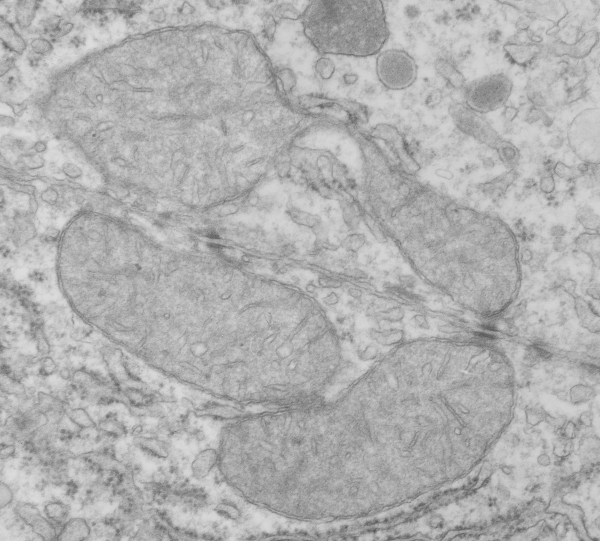
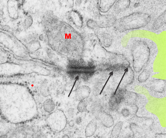
THis Electron micrograph has at least two mitochondria that are attached by intermediate filaments to desomosomal junctions. Adjacent hepatocytes from a rat, which was exposed to carbon monoxide while pregnant with pups. I have to presume this biopsy was taken at the time pups were delivered, around GD 21. Incidentally there are some funny mitochondrial shapes here, and lots of vesicles, and peroxisosomes. I do not remember if any morphometry was performed on these liver samples.
Daily Archives: January 11, 2018
Desmosomal mitochondrial tethers: fetal follicular cells of the thyroid
Just in case anyone is interested in whether there are desmosomes which are tethered to mitochondria via intermediate filaments in cells other than those I have reported so far then look at a fetal follicular cell (of the thyroid) and it is very apparent that these pretty large desmosomes with mitochondrial attachment via filaments is certainly happening. This site is by the tight junction, adherens junction, and the desmosome. Just like hepatocyte D-M tethers, which tend to occur around bile canaliculi in hepatocytes.
Red M, mitochondrion; three arrows point to three different types of intercellular junctional complexes (including the desmosome and the mitochondrion (most left of the three); colloid in the lumen (light green shading) can be seen on the right hand side of the electron micrograph. Red dot is @ 27nm in diameter representing the size of a ribosome.