Monthly Archives: December 2017

Desmosomes in alveolar type II cells: perhaps not? or if so, not very many!

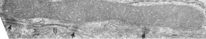
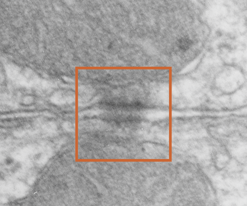
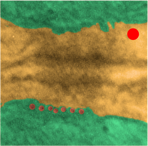
While perusing hundreds of electron micrographs of alveolar type II cells in half a dozen species i was disappointed NOT to see any desmosomes, or if I saw them they were not very well developed and tangentially cut, but I really think they may exist in a different form than they do, at least, in liver.

This I surmise from the fact that very little of the alveolar type II cell is actually “tethered” to other cells. A large portion (basal plasmalemma) abuts basement membrane, a tiny bit is close to alveolar type I cells, that connection by the way is very thin and would not even provide enough space wherein a mitochondria could reside and be found tethered to a desmosome, if i could find a desmosome. The large (perhaps the largest) plasmalemmal surface is in the alveolar space and no desmosomes possible there.  So the hunt for desmosomal mitochondrial tethers in alveolar type II cells just came up empty. Empty for desmosomes, empty for desmosomal mitochondrial tethers, but of course there are plenty of mitochondria per se…. just not tethered to anything resembling a desmosome.  I don’t even have an image of something which might be considered “close”.

It makes sense in terms of physiology however not to have rigid structures tightly binging alveolar type II cells together…. the tremendous movement within the alveolus durin inspiration and expiration would make it a little untenable to have to use energy, move mitochondria, make and break associations within the cell and the intermediate filaments, and lose the intercellular bonds in the cadherin molecules 20 or 30 times a minute. (and remember that is slow compared to the inspiration and expiration of some animals).  So peripheral lung was probably a silly tissue to hunt for desmosomal mitochondrial tethers (there are other junctional complexes of course to prevent fluid from coming into the intercellular space that are easily seen, tight junctions and adherens junctions. Some describers of peripheral lung epi dont even mention desmosomes. HERE (i have never heard of this journal, so reader beware)

Anticipatory Anxiety – Adrenaline Addicts: a sad commentary

Stress lets us know we are alive. When we need stimulation because we haven’t had enough exercise, or when we are in physical danger (or even just hungry or hurrying up to get somewhere) we pump out adrenaline.  This signals to our brains…. wow, we ARE really alive.  When we are bored or are feeling under-stimulated, we purposefully do things (un- or subconsciously or even overtly) to shoot adrenaline.  Being addicted to adrenaline (you’ve heard of it, the adrenaline junkie, or maybe the adrenaline addict) is something which we have become in our society, and are continually becoming to greater and greater degrees. You have certainly heard the mantras “I’ve been worried sick” or “I was worried to death”.  Those old guys knew from astute observations, what we have forgotten. Worrying does kill.

Fuel for becoming an AA – is fed by our taste for social media, rapidly moving TV and movie thrillers, global news, our own rage or just worrying (anticipating) things we think might happen.  We are a world of Adrenaline Addicts. “The most concrete function attributed to worry is an attempt at constructive mental problem solving, although it may represent an unproductive and, perhaps, even a counterproductive attempt”

The sequelae?  shorter life, more metabolic diseases, waist-line fat, diabetes, vascular diseases, depression — and more — certainly that is not the abundant life.
(CORTISOL from wikipedia commons)Cortisol-3D-balls

Kroger frozen chopped broccoli-

I like the convenience of frozen food. I am willing to admit that I don’t buy organic (too many questions about the rules, allowances and processes for what makes something organic dont add up) and I am not sure that the issue i am about to explain would be covered there anyway. I do get tired of poor quality control.  I am not able to judge the amount of pesticide residue left in frozen foods by lazy and inadequate preparation techniques…. that bothers me, but it is not something i can taste or “see” or “not chew”. What I dont like is having inedible pieces of brocolli stem in my food.

I get tired of having to call Kroger and tell them that they need to change something only to hear the “we are sorry for any inconvenience” shpieel, and have them fake-plakate me by sending a coupon.  I would really like them to be attentive and to make changes where appropriate.

Today’s issue has gone on for months and months. I logged in a call about the chopped broccoli (see picture) many many months ago and gave the UPC  code along with the number of packages of frozen broccoli I had purchased with pieces of very tough stem throughout the bag. The problem is with the vendor…. they are chopping up broccoli, yes, but so far down the stem of the broccoli that there are pieces in with the chopped broccoli that are totally inedible.  Inedible, tough, and that chew like wood.

Trimming off a little more of the stem obviously will cut into their supplier’s profit, but that would pale in comparison to individuals avoiding the product altogether. That is not a big change to make…  but here it is six months later and i am still finding yellow-white broccoli woody stems in my broccoli.  Check out your own bags of Kroger frozen chopped broccoli and see if i am right. Then call them and complain.

What element brings together intermediate filaments, mitochondria and desmosomes?

What element brings together intermediate filaments, mitochondria and desmosomes? I think it would be nice to know which of these organelles is responsible for initiating the association of the three to create the desmosomal mitochondria tether which is connected via intermediate filaments. Which?
1) Intermediate filaments can “tug” on mitochondrial shape (morphology). This is clearly evident in countless micrographs.
2) Intermediate filaments bind with the desmoplakin protein (part of the desmosome) and the latter bonds with the dense portion of the desmosome (plakophilin, plakoglobin) just adjacent to the plasmalemma (on the intracellular side) and the intracellular ends of desmocolin and desmoglein (the main proteins in the intercellular space).
3) Mitochondria as calcium buffers may have be important in regulating the calcium dependent adhesion molecules, desmocolin and desmoglein, in particular formation and reduction of desmosomes. This is a function seen in other intermediate filament mitochondria tethers as well.
4) Mitochondria provide energy as ATP.
5) Also, the flattened part of the mitochondrial membrane which is adjacent to the desmosomal – mitochondrial tether area with intermediate filaments would probably include mitochondrial membrane anchoring receptors.
6) If what is reported is true (longer mitochondria produce more energy) then those long three or four thered areas on a single mitochondria would be “really active” in terms of energy production). Arrowheads = des – mit tethers; tj = tight junction; bar = 100nm; RER along the top right of the image. Syrian hamster hepatocyte.
A LIST OF VARIOUS accepted functions for membrane-mitochondria-filament associations
a- apoptosis
b- lipid transport
c- autophagy
d- mitochondrial morphology
e- intercellular junctions
So this is a obviously going to be the short list…. thee will be many more functions to come.

Desmosomal-mitochondrial tethers: x2

Here is a gallery sheet of desmosomal mitochondrial tethers, and some are double, some are double-doubles (haha) and some have three tethers.  I can’t overemphasize how interesting these little structures are and how critical they must be for cell function. Just considering how the desmosome itself has become a known mechanism of cell signaling, then the energy provided (mitochondria) and the movement of proteins and mitochondria and signaling proteins via the intermediate filaments, and the latter involved also in cell shape change…. makes these structures very very important.  This gallery (making the gallery itself) caused me great grief… ha ha.. my little desk top computer just didn’t want to handle composite images of over a gig.  Would that I had real equipment…  wonder of wonders.  Enjoy the image(s) of whatever these DES-MiTes  (desmosomal-mitochondrial-tethers)  will ultimately be called (dumber names exist in science).

34 pix, 43 desmosomes, at least 56 mitochondria, numerous species represented here (ferret, rat, guinea pig, stub tail monkey, rhesus monkey, maybe others, and all images are from hepatocyte-hepatocyte junctions)

desmosomal mitochondria tethers and double tethers in hepatocytes


A time for Christmas

Christmas is an odd mix of pagan traditions and real live love, sacrifice and hope that the real PEACE reigns…. Sometimes coming to grips with both is a little taxing for the frontal lobe. Here is my “resolution” ha ha.

Questions about desmosomes and mitochondrial junctions

Desmosomes:

(who names these proteins — desmosome..  desmo — and soma, that is the easy button, cadherins…  well calcium dependent adhering molecules, i guess thats ok, desmoglobins… not so good, plakophilin, maybe that should be desmoplakoglobofilin.. ha ha…  i guess too that the plaque, and plako…. go together, but there are two intracellular zones (apparently) of the desmosome…  inner (which is already confusing because to me, if the desomosome is the unit being described, then inner would be the closest plaque to the center of the desmosome, which is the central dense line in the intercellular space, but no…someone  named it “backwards” with the center of the CELL rather than the center of the DESMOSOME as the reference point, thus what is named the outer desmosomal plaque is actually “inner” with reference to the desmosome).  It is astounding how differently we all describe our world– and it adds a whole lot of confusion and difficulty to learning and understanding).

More or less facts:
1) electron dense disc like plaques (Stedman’s medical dictionary recommends the spelling “disc” for all medical uses) so I will use disc.
2) extracellular-intra-and-intercellular-cell-to-cell adhesion discs
3) prominent in epithelial cells
4) 0.2 to 0.5 microns in the flat dimension (depending likely on whether the edge of the disc is in the plane of section or the center
5) very likely different states of cell activity change the dimension and the number and the location of desmosomes as well as the number of mitochondria which are associated.
6) Only a small percent of mitochondria are associated with desmosomes, mainly because there are many mitochondria in each cell. (Volume vs perimeter consideration).

Desmosomal cadherins – desmocolin (3 isoforms) and desmoglein (4 or more isoforms). DSC2 and DSG2 are the most common.

Tying the desmosome to intercellular elements are the “intermediate filaments: a diverse class of flexible filaments that provide mechanical strength to cells. They make up hair, nail, horn, and scale cells, they form the nuclear lamina, which lies just inside the inner nuclear membrane, they span cells to provide strength to epithelial tissues, and they anchor organelles and stabilize the cytoplasm” with these data (Intermediate filaments 6–10 strands per filament (6 was quoted for those which are next to desmosomes), non-polar, outside diameter he quotes as ~10 nm (I think this might be larger, or maybe stains add dimensions)) an edited quote from Methods for modeling cytoskeletal and DNA filaments by Steven Andrews (Physical Biology, Volume 11, Number 1 )

Desmoglein 2

“Desmoglein-2 is a 122.2 kDa protein composed of 1118 amino acids. Desmoglein-2 is a calcium-binding transmembrane glycoprotein component of desmosomes in vertebrate cells. Currently, four desmoglein subfamily members have been identified and all are members of the cadherin cell adhesion molecule superfamily.”

Intermediate filaments – desmosomes – mitochondria

I havn’t read much (or found much to read) about the making – breaking – moving – adjusting of desmosomes, or the energy requirement for desmosome modification, but it seems too much a coincidence that mitochondria would be closely tethered to desmosomes and not provide this function. That said, they have been photographed by me in mouse, rat, neonatal rat, ferret, hamster, syrian hamster, dog, and guinea pig, just to name a few in the list that I have looked at in the last weeks. And they are so similar in all, that so far, if i list one image of a desmosomal-mitochondrial junction from one species I cannot tell it from another. This is good, i guess, but it would be more interesting if the structure showed some variation in structure….. that could perhaps be correlated with differences in the component proteins.

Here is one desmosomal-mitochondrial junction from ferret, which didn’t have a nice even pattern of cadherins in the middle, but did have a punctuated central line…with dots less than 20nm apart…. they seemed a little staggered.  Here is the original micrograph (i removed a scratch in the negative not in the area of the desmosome using photoshop bandaid tool, but it otherwise just a scan of the print. 5940 18648 ferret hepatocyte.

The image below it is an enlargement of the box in top image. Ribosome-red dot for size, and the black lines overlie densities in the intercellular area of the desmosome.


Desmosome-to-mitochondria: connections

More desmosomal mitochondria tethering from hamster hepatocytes. This particular view has two mitochondria, and one shows a line of “dots” or fibers or fibrils in cross section.

This image is from a syrian hamster hepatocyte, and it is just a routine TEM but I noticed (while making my gallery collection of desmosomal-mitochondrial images) that one of the mitochondria showed the repeating dots (cross sections likely of intermediate filaments (maybe types I and II).

Upper image is unretouched micrograph and lower image has been pseudocolored. The latter has green for the two mitochondria that are tethered to the desmosome  at their outer mitochondria membrane (which appears rigid where intermediate filaments are present, and a distinct and definitely separated inner mitochondrial membrane). Along with the intermediate filaments are connections to the desmoplakin molecules the latter being part of the outer dense plaque of the desmosome. The yellow is the cytoplasmic area of the two adjacent cells including the intercellular space and the spot desmosome. Red dot is appoximately the size of a ribosome (27nm) which makes these filaments cut in cross section about 13.5nm in diameter (orange dots in a row) bottom electron micrograph. This hastily made measurement is not that far off from the reported 10nm intermediate filament made up of four monomer filament tetramers (haha… figure that out).

According to Thomason et al,  desomosomal proteins are as follows (slightly rearranged to make more sense to me): Desmosomes have five major component proteins, the DCs (desmosomal cadherins) DSG (desmoglein) and DSC (desmocollin) –DSC and DSG are the desmosomal adhesion molecules. Plakin family cytolinker DP (desmoplakin), and the arm (armadillo) proteins PG (plakoglobin) and PKP (plakophilin).  DP links the desmosomal plaque to the IF (intermediate filament) cytoskeleton, and PG and PKP are adaptor proteins that link between the adhesion molecules and DP.