Monthly Archives: February 2018

Early article on desmosomal-mitochondrial complexes

After so many years of looking for articles on this topic (I actually wondered how this phenomenon could go unnoticed for so many decades) i did find one manuscript (which references two others) by Deane et al 1966 which mentions them and shows several micrographs (sometimes pretty low manification). They found these first in fetal and neonatal tissues, so this is a clue and also important for the concept that I have which is that in tissues with higher rates of cell division that also are epithelia which connect at environmental interfaces (meaning the “outside” whether that is physically outside or not) have an active mechanism for building and disassembling desmosomes.  They felt it was a one-way passage…. i think it is more a modeling-remodeling phenomenon.  So this was nice but bitter sweet at the same time since I thought my early paper in a very obscure journal in the 1980s was the first.

(I am going to use this acronym…desmosomal mitochondrial tethers  regardless of how silly it sounds just because it is too time consuming to type out 32 characters when 7 will do, my apologies.)

miller et al, desmosomal mitochodnrial interactions_1985
deane_1966_desmosomal mitochondrial complexes
In figure 1, and enlargement inset called figure 2 (a portion of which is below) of their article, they make a cute comment…. about a desmosome beginning to develop nearby to a reasonably well defined desmite. It would be better perhaps to have recognized that there were two possibilities — one, that it was a tangential cut to a sequentially placed tether along a single mitochondrial outer membrane, and the other that it was a desmite “being built or broken”. This little area of not completely well defined junction they label with a black arrow. You can see that it is not quite a desmosome and the mitochondrion really isn’t that close to it.  The label TF (i don’t know why it is in italics) surely stands for tonofilaments…. which I refer to as intermediate filaments.
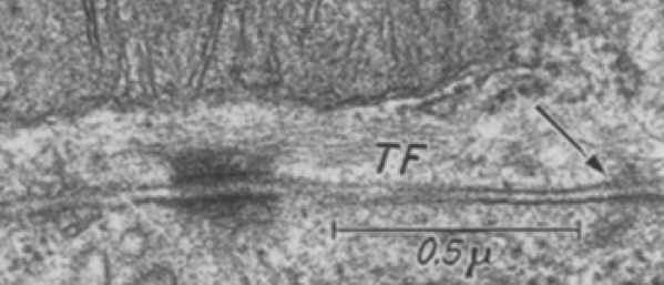

Just using their image and their micron marker (0.5mu = 500nm) it seems that their cut in this desmosome is just off center but close (diameter with the annulus) at about 286nm (central cut yields a desmosome of 300nm) and a diameter of 214nm excluding the annulus.  About 162nm span the distance from the outer mitochondrial membrane to the plasmalemma membrane of the desmosome of the cell on the same side. A quick measurement for the thickness of the extracellular space of the desmosome in this image was about 18nm.  About 15nm /2 for the dimension of the annulus area on either side of the desmosomal spot. This micrograph shows unequal annulus dimensions.  Lovingly I point out the scratch (likely on the old acetate negative) lower left side of the desmosome… and in sympathy say, my negatives and micrographs have many such scratches. One thing to not is that 3-5 intermediate filaments are lying almost parallel to the plasmalemma and the outer mitochondrial membrane… certainly NOT like the diagrams seen routinely  — see my post with desmosomal diagrams with hairpin (wrongly directed) lines for intermediate filament attachments to desmoplakin molecules.
What we don’t know we either fear or worship!

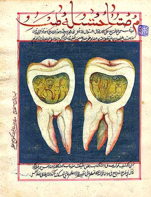
What we don’t know we either hate or worship! How strange this is, yet for eons this has been true. I was making a crossword puzzle, using the concept of heaven and how oddly this place-space-destination-resting spot-culmination-equilizer-paradise concept has changed over time. I ran onto the concept of JNN or jinn or jinni or anglicized as genie, and found this gorgeous page from a manuscript.  The jinn were important god-like beings at one point near pre-history but were demoted to human like then smaller and smaller unknown forces for events and ailments that were not understood.  All dentists will totally love this image of two teeth, with the pulp and root canals occupied by jinn….  causing a tooth ache.

As soon as the elements causing teeth to rot and die and ache, jinn went on to become some other force, no longer hated nor worshiped.  We are a strange species at best. Thank you wikipedia.


Desmosomal symmetry: not a complete mirror images side to side

Desmosomal symmetry: not a complete mirror images side to side, since it seems to occur in my micrographs, and obviously too in this micrograph from Green and Gaudry, that the separate cells (adjacent cells bound by the single desmosome) may have a propensity to have intermediate filaments coursing by at perpendicular angles.  Maybe also chance – I guess it is a 50 50 chance to be chance (haha).

But also in this quote from Green and Gaudry.…I take exception to their remark that the desmosome is tripartite. A quote from their paper yielded this text of which several parts make no sense to me “electron micrograph further illustrates the highly organized ultrastructure of a desmosome (yes, i agree) in which mirror-image (partly agree, but not always true), tripartite electron-dense plaques (show me 3, ha ha, and are you counting lucent bands, plasmalemma (not really to be counted and intermediate filaments?) sandwich a central core consisting of adjacent plasma membranes (wait, plasmalemmas are NOT part of the central CORE, and what is meant by CORE anyway, that is a term that needs definition) bisected by an intercellular zipper-like midline (yes, zipper like dense midline -i agree)

They didn’t take into acount the different directions (which may be purposeful) of the intermediate filaments coursing by the desmoplakin molecules in the adjacent cells… as sometimes they appear as 10 nm cross sections and other times as low arcing swooshes. I ask, what part of the plaques are divided into 3??? This is a confusing, since there is nothing distinct about the 3 parts they refer to… they could represent many/or any different layers of this organized structure. Their own electron micrograph (pasted below) shows pretty convincingly that there is not total mirror symmetry to the desmosome since their cell on the bottom part of the image shows cross sections of intermediate filaments while the cell on the top part of their image shows the longitudinal swoosh of intermediate filaments.

One thing that their electron micrograph shows that is rarely commented on is the desmosome annulus… this ring which of plasmalemma which is just slightly morphologically different than plasmalemma further from the desmosome.

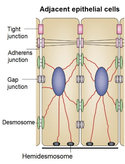
While on the topic of their diagram, i think more care could have been extended to the depiction of the tight junction which is really a weld, and they show it as a wider structure than the two blended plasmalemmal membranes really are. And the adherens junctions are diagrammed to be as prominent as desmosomes, which they are not, and the labeling of adherens junctions actually appears closer to their green blob desmosome and thus doesn’t really direct attention to the junction they are wanting to show.  Maybe that works for a visual aid for some, but for me it does the opposite…. working from a place of knowing the actual structures and trying to figure out what visual errors the “diagrammer” has created… is frustrating.  This brings up the legion of diagrams of really poorly drawn desmosomal structures… ha ha.  I do wish more care could be taken in scientific illustrations..N
Not a single diagram that I have seen on desmosomes and intermediate filaments has mentioned the importance of the presence of mitochondria in building and breaking down these structures.